 ムービー
ムービー コントローラー


+ データを開く

- 基本情報
基本情報
| 登録情報 | データベース: PDB / ID: 5sha | ||||||
|---|---|---|---|---|---|---|---|
| タイトル | CRYSTAL STRUCTURE OF HUMAN PHOSPHODIESTERASE 10 IN COMPLEX WITH n2c(c1nc(nn1c(c2)C)CCc3nc(nn3C)N4C[C@@H](C(F)F)CC4)C, micromolar IC50=0.048101 | ||||||
 要素 要素 | cAMP and cAMP-inhibited cGMP 3',5'-cyclic phosphodiesterase 10A | ||||||
 キーワード キーワード | HYDROLASE/HYDROLASE inhibitor / PHOSPHODIESTERASE / PDE10 / HYDROLASE / SCHIZOPHRENIA / HYDROLASE-HYDROLASE inhibitor complex | ||||||
| 機能・相同性 |  機能・相同性情報 機能・相同性情報3',5'-cGMP-stimulated cyclic-nucleotide phosphodiesterase activity / 3',5'-cyclic-nucleotide phosphodiesterase / negative regulation of receptor guanylyl cyclase signaling pathway / cGMP catabolic process / cAMP catabolic process / regulation of cAMP/PKA signal transduction / cGMP effects / 3',5'-cyclic-nucleotide phosphodiesterase activity / regulation of adenylate cyclase-activating G protein-coupled receptor signaling pathway / cGMP binding ...3',5'-cGMP-stimulated cyclic-nucleotide phosphodiesterase activity / 3',5'-cyclic-nucleotide phosphodiesterase / negative regulation of receptor guanylyl cyclase signaling pathway / cGMP catabolic process / cAMP catabolic process / regulation of cAMP/PKA signal transduction / cGMP effects / 3',5'-cyclic-nucleotide phosphodiesterase activity / regulation of adenylate cyclase-activating G protein-coupled receptor signaling pathway / cGMP binding / 3',5'-cyclic-GMP phosphodiesterase activity / 3',5'-cyclic-AMP phosphodiesterase activity / : / cAMP binding / G alpha (s) signalling events / glutamatergic synapse / metal ion binding / cytosol 類似検索 - 分子機能 | ||||||
| 生物種 |  Homo sapiens (ヒト) Homo sapiens (ヒト) | ||||||
| 手法 |  X線回折 / シンクロトロン / 分子置換 / 解像度: 2.24 Å X線回折 / シンクロトロン / 分子置換 / 解像度: 2.24 Å | ||||||
 データ登録者 データ登録者 | Joseph, C. / Benz, J. / Flohr, A. / Rudolph, M.G. | ||||||
| 資金援助 |  スイス, 1件 スイス, 1件
| ||||||
 引用 引用 | ジャーナル: To be published タイトル: Crystal Structure of a human phosphodiesterase 10 complex 著者: Flohr, A. / Schlatter, D. / Kuhn, B. / Rudolph, M.G. | ||||||
| 履歴 |
|
- 構造の表示
構造の表示
| 構造ビューア | 分子: MolmilJmol/JSmol |
|---|
- ダウンロードとリンク
-ダウンロード
| PDBx/mmCIF形式 | 5sha.cif.gz | 274.1 KB | 表示 | PDBx/mmCIF形式 |
|---|---|---|---|---|
| PDB形式 | pdb5sha.ent.gz | 220.7 KB | 表示 | PDB形式 |
| PDBx/mmJSON形式 | 5sha.json.gz | ツリー表示 | PDBx/mmJSON形式 | |
| その他 |  その他のダウンロード その他のダウンロード |
-検証レポート
| 文書・要旨 | 5sha_validation.pdf.gz | 1.3 MB | 表示 | wwPDB検証レポート |
|---|---|---|---|---|
| 文書・詳細版 | 5sha_full_validation.pdf.gz | 1.3 MB | 表示 | |
| XML形式データ | 5sha_validation.xml.gz | 55.8 KB | 表示 | |
| CIF形式データ | 5sha_validation.cif.gz | 73.8 KB | 表示 | |
| アーカイブディレクトリ | https://data.pdbj.org/pub/pdb/validation_reports/sh/5shaftp://data.pdbj.org/pub/pdb/validation_reports/sh/5sha | HTTPS FTP |
-グループ登録
| ID | G_1002229 (175エントリ) |
|---|---|
| タイトル | To be published |
| タイプ | undefined |
| 解説 | A set of PDE10 crystal structures |


-関連構造データ
| 類似構造データ |
|---|
-リンク
 PDBj
PDBj

- 集合体
集合体
| 登録構造単位 | 
| ||||||||
|---|---|---|---|---|---|---|---|---|---|
| 1 | 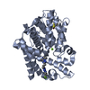
| ||||||||
| 2 | 
| ||||||||
| 3 | 
| ||||||||
| 4 | 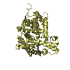
| ||||||||
| 単位格子 |
|
-要素
| #1: タンパク質 | 分子量: 39413.203 Da / 分子数: 4 / 由来タイプ: 組換発現 / 由来: (組換発現) Homo sapiens (ヒト) / 遺伝子: PDE10A / プラスミド: PET28a(+) / 発現宿主:  参照: UniProt: Q9Y233, 3',5'-cyclic-nucleotide phosphodiesterase #2: 化合物 | ChemComp-ZN /   分子量: 65.409 Da / 分子数: 4 / 由来タイプ: 合成 / 式: Zn 分子量: 65.409 Da / 分子数: 4 / 由来タイプ: 合成 / 式: Zn#3: 化合物 | ChemComp-MG /   分子量: 24.305 Da / 分子数: 4 / 由来タイプ: 合成 / 式: Mg 分子量: 24.305 Da / 分子数: 4 / 由来タイプ: 合成 / 式: Mg#4: 化合物 | ChemComp-JBV / (   分子量: 376.407 Da / 分子数: 4 / 由来タイプ: 合成 / 式: C17H22F2N8 / タイプ: SUBJECT OF INVESTIGATION 分子量: 376.407 Da / 分子数: 4 / 由来タイプ: 合成 / 式: C17H22F2N8 / タイプ: SUBJECT OF INVESTIGATION#5: 水 | ChemComp-HOH / |  分子量: 18.015 Da / 分子数: 406 / 由来タイプ: 天然 / 式: H2O 分子量: 18.015 Da / 分子数: 406 / 由来タイプ: 天然 / 式: H2O研究の焦点であるリガンドがあるか | Y | Has protein modification | Y | |
|---|
-実験情報
-実験
| 実験 | 手法: X線回折 / 使用した結晶の数: 1 |
|---|
- 試料調製
試料調製
| 結晶 | マシュー密度: 2.6 Å3/Da / 溶媒含有率: 52.73 % |
|---|---|
| 結晶化 | 温度: 295 K / 手法: 蒸気拡散法, シッティングドロップ法 / pH: 7.5 詳細: 5-20 mg/mL protein in 25mM HEPES/NaOH pH7.5, 150mM NaCl, 50mM BME mixed 1:1 with reservoir 0.1M HEPES/NaOH pH7.5, 30% PEG550MME, 50mM MgCl2 |
-データ収集
| 回折 | 平均測定温度: 100 K | ||||||||||||||||||||||||||||||||||||||||||||||||||||||||||||||||||||||||||||||||||||||||||||||||||||||||||||||||||||||||||||||||||||||||||||||||||||||||||||||||||||||||||||||||||||||||||||||||||||||||||||||||||
|---|---|---|---|---|---|---|---|---|---|---|---|---|---|---|---|---|---|---|---|---|---|---|---|---|---|---|---|---|---|---|---|---|---|---|---|---|---|---|---|---|---|---|---|---|---|---|---|---|---|---|---|---|---|---|---|---|---|---|---|---|---|---|---|---|---|---|---|---|---|---|---|---|---|---|---|---|---|---|---|---|---|---|---|---|---|---|---|---|---|---|---|---|---|---|---|---|---|---|---|---|---|---|---|---|---|---|---|---|---|---|---|---|---|---|---|---|---|---|---|---|---|---|---|---|---|---|---|---|---|---|---|---|---|---|---|---|---|---|---|---|---|---|---|---|---|---|---|---|---|---|---|---|---|---|---|---|---|---|---|---|---|---|---|---|---|---|---|---|---|---|---|---|---|---|---|---|---|---|---|---|---|---|---|---|---|---|---|---|---|---|---|---|---|---|---|---|---|---|---|---|---|---|---|---|---|---|---|---|---|---|---|
| 放射光源 | 由来: シンクロトロン / サイト: SLS / ビームライン: X10SA / 波長: 1 Å | ||||||||||||||||||||||||||||||||||||||||||||||||||||||||||||||||||||||||||||||||||||||||||||||||||||||||||||||||||||||||||||||||||||||||||||||||||||||||||||||||||||||||||||||||||||||||||||||||||||||||||||||||||
| 検出器 | タイプ: PSI PILATUS 6M / 検出器: PIXEL / 日付: 2013年3月17日 | ||||||||||||||||||||||||||||||||||||||||||||||||||||||||||||||||||||||||||||||||||||||||||||||||||||||||||||||||||||||||||||||||||||||||||||||||||||||||||||||||||||||||||||||||||||||||||||||||||||||||||||||||||
| 放射 | プロトコル: SINGLE WAVELENGTH / 単色(M)・ラウエ(L): M / 散乱光タイプ: x-ray | ||||||||||||||||||||||||||||||||||||||||||||||||||||||||||||||||||||||||||||||||||||||||||||||||||||||||||||||||||||||||||||||||||||||||||||||||||||||||||||||||||||||||||||||||||||||||||||||||||||||||||||||||||
| 放射波長 | 波長: 1 Å / 相対比: 1 | ||||||||||||||||||||||||||||||||||||||||||||||||||||||||||||||||||||||||||||||||||||||||||||||||||||||||||||||||||||||||||||||||||||||||||||||||||||||||||||||||||||||||||||||||||||||||||||||||||||||||||||||||||
| 反射 | 解像度: 2.24→43.32 Å / Num. obs: 76396 / % possible obs: 99.9 % / 冗長度: 5.186 % / Biso Wilson estimate: 52.472 Å2 / CC1/2: 0.999 / Rmerge(I) obs: 0.082 / Rrim(I) all: 0.091 / Χ2: 0.848 / Net I/σ(I): 15.1 / Num. measured all: 396216 / Scaling rejects: 43 | ||||||||||||||||||||||||||||||||||||||||||||||||||||||||||||||||||||||||||||||||||||||||||||||||||||||||||||||||||||||||||||||||||||||||||||||||||||||||||||||||||||||||||||||||||||||||||||||||||||||||||||||||||
| 反射 シェル | Diffraction-ID: 1
|
- 解析
解析
| ソフトウェア |
| |||||||||||||||||||||||||||||||||||||||||||||||||||||||||||||||||||||||||||
|---|---|---|---|---|---|---|---|---|---|---|---|---|---|---|---|---|---|---|---|---|---|---|---|---|---|---|---|---|---|---|---|---|---|---|---|---|---|---|---|---|---|---|---|---|---|---|---|---|---|---|---|---|---|---|---|---|---|---|---|---|---|---|---|---|---|---|---|---|---|---|---|---|---|---|---|---|
| 精密化 | 構造決定の手法: 分子置換 開始モデル: inhouse model 解像度: 2.24→43.32 Å / Cor.coef. Fo:Fc: 0.97 / Cor.coef. Fo:Fc free: 0.946 / SU B: 7.577 / SU ML: 0.173 / 交差検証法: THROUGHOUT / σ(F): 0 / ESU R: 0.247 / ESU R Free: 0.204 / 立体化学のターゲット値: MAXIMUM LIKELIHOOD 詳細: difluoromethyl clashes with nearby Glu, and in some molecules also Pro. Has different conformations and appears more flexible than rest of ligand based on B-values.
| |||||||||||||||||||||||||||||||||||||||||||||||||||||||||||||||||||||||||||
| 溶媒の処理 | イオンプローブ半径: 0.8 Å / 減衰半径: 0.8 Å / VDWプローブ半径: 1.2 Å / 溶媒モデル: MASK | |||||||||||||||||||||||||||||||||||||||||||||||||||||||||||||||||||||||||||
| 原子変位パラメータ | Biso max: 136.42 Å2 / Biso mean: 46.608 Å2 / Biso min: 16.96 Å2
| |||||||||||||||||||||||||||||||||||||||||||||||||||||||||||||||||||||||||||
| 精密化ステップ | サイクル: final / 解像度: 2.24→43.32 Å
| |||||||||||||||||||||||||||||||||||||||||||||||||||||||||||||||||||||||||||
| 拘束条件 |
| |||||||||||||||||||||||||||||||||||||||||||||||||||||||||||||||||||||||||||
| LS精密化 シェル | 解像度: 2.24→2.298 Å / Rfactor Rfree error: 0 / Total num. of bins used: 20
|
