positive regulation of chromatin binding / [histone H3]-trimethyl-L-lysine9 demethylase / histone H3K9me2/H3K9me3 demethylase activity / positive regulation of double-strand break repair via nonhomologous end joining / histone H3K9 demethylase activity / regulation of protein phosphorylation / histone demethylase activity / pericentric heterochromatin / cellular response to ionizing radiation / HDMs demethylate histones ...positive regulation of chromatin binding / [histone H3]-trimethyl-L-lysine9 demethylase / histone H3K9me2/H3K9me3 demethylase activity / positive regulation of double-strand break repair via nonhomologous end joining / histone H3K9 demethylase activity / regulation of protein phosphorylation / histone demethylase activity / pericentric heterochromatin / cellular response to ionizing radiation / HDMs demethylate histones / chromatin DNA binding / double-strand break repair via homologous recombination / site of double-strand break / regulation of gene expression / blood microparticle / damaged DNA binding / chromatin remodeling / inflammatory response / chromatin / nucleoplasm / metal ion binding / nucleus Similarity search - Function
JmjN domain / jmjN domain / JmjN domain profile. / Small domain found in the jumonji family of transcription factors / Cupin / JmjC domain, hydroxylase / A domain family that is part of the cupin metalloenzyme superfamily. / JmjC domain / JmjC domain profile. / Jelly Rolls ...JmjN domain / jmjN domain / JmjN domain profile. / Small domain found in the jumonji family of transcription factors / Cupin / JmjC domain, hydroxylase / A domain family that is part of the cupin metalloenzyme superfamily. / JmjC domain / JmjC domain profile. / Jelly Rolls / Sandwich / Mainly Beta Similarity search - Domain/homology
PanDDA analysis group deposition of models of ground state datasets
Type
ground state
Description
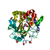
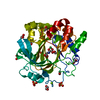
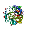
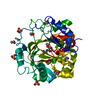
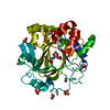
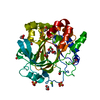
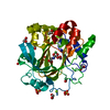
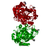
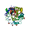
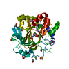
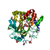
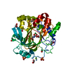
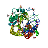
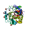
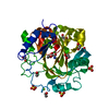
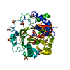
Jmjc domain of human JMJD2D screened against the ZENOBIA Fragment Library by X-ray Crystallography. Check out the PanDDA event maps at https://zenodo.org/record/290220/files/0_index.html
Lysine-specificdemethylase4D / JmjC domain-containing histone demethylation protein 3D / Jumonji domain-containing protein 2D
Mass: 42050.539 Da / Num. of mol.: 1 Source method: isolated from a genetically manipulated source Source: (gene. exp.) Homo sapiens (human) / Gene: KDM4D, JHDM3D, JMJD2D / Production host: Escherichia coli (E. coli) References: UniProt: Q6B0I6, Oxidoreductases; Acting on paired donors, with incorporation or reduction of molecular oxygen; With 2-oxoglutarate as one donor, and incorporation of one atom of oxygen into each donor
In the structure databanks used in Yorodumi, some data are registered as the other names, "COVID-19 virus" and "2019-nCoV". Here are the details of the virus and the list of structure data.
Jan 31, 2019. EMDB accession codes are about to change! (news from PDBe EMDB page)
EMDB accession codes are about to change! (news from PDBe EMDB page)
The allocation of 4 digits for EMDB accession codes will soon come to an end. Whilst these codes will remain in use, new EMDB accession codes will include an additional digit and will expand incrementally as the available range of codes is exhausted. The current 4-digit format prefixed with “EMD-” (i.e. EMD-XXXX) will advance to a 5-digit format (i.e. EMD-XXXXX), and so on. It is currently estimated that the 4-digit codes will be depleted around Spring 2019, at which point the 5-digit format will come into force.
The EM Navigator/Yorodumi systems omit the EMD- prefix.
Related info.:Q: What is EMD? / ID/Accession-code notation in Yorodumi/EM Navigator
Yorodumi is a browser for structure data from EMDB, PDB, SASBDB, etc.
This page is also the successor to EM Navigator detail page, and also detail information page/front-end page for Omokage search.
The word "yorodu" (or yorozu) is an old Japanese word meaning "ten thousand". "mi" (miru) is to see.
Related info.:EMDB / PDB / SASBDB / Comparison of 3 databanks / Yorodumi Search / Aug 31, 2016. New EM Navigator & Yorodumi / Yorodumi Papers / Jmol/JSmol / Function and homology information / Changes in new EM Navigator and Yorodumi
 Movie
Movie Controller

 Yorodumi
 Open data
 Basic information
 Components
 Keywords
 Function and homology information
 Homo sapiens (human)
 X-RAY DIFFRACTION /
  Authors
 Citation
 Structure visualization
 Downloads & links
 Other downloads
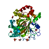


 PDBj
PDBj
 Assembly


 Mass: 65.409 Da / Num. of mol.: 1 / Source method: obtained synthetically / Formula: Zn
 Mass: 58.693 Da / Num. of mol.: 1 / Source method: obtained synthetically / Formula: Ni
 Mass: 24.305 Da / Num. of mol.: 1 / Source method: obtained synthetically / Formula: Mg
 Mass: 147.086 Da / Num. of mol.: 1 / Source method: obtained synthetically / Formula: C4H5NO5 / Comment: inhibitor*YM
 Mass: 62.068 Da / Num. of mol.: 8 / Source method: obtained synthetically / Formula: C2H6O2
 Mass: 96.063 Da / Num. of mol.: 3 / Source method: obtained synthetically / Formula: SO4
 Sample preparation
 / Beamline: I03 / Wavelength: 0.9763 Å
 Processing