Initiation of Nuclear Envelope (NE) Reformation / regulation of protein desumoylation / regulation of nucleocytoplasmic transport / nuclear pore central transport channel / transcription-dependent tethering of RNA polymerase II gene DNA at nuclear periphery / nuclear pore complex assembly / phosphatidylcholine biosynthetic process / import into nucleus / Transport of Mature mRNA derived from an Intron-Containing Transcript / Regulation of HSF1-mediated heat shock response ...Initiation of Nuclear Envelope (NE) Reformation / regulation of protein desumoylation / regulation of nucleocytoplasmic transport / nuclear pore central transport channel / transcription-dependent tethering of RNA polymerase II gene DNA at nuclear periphery / nuclear pore complex assembly / phosphatidylcholine biosynthetic process / import into nucleus / Transport of Mature mRNA derived from an Intron-Containing Transcript / Regulation of HSF1-mediated heat shock response / nuclear pore nuclear basket / SUMOylation of SUMOylation proteins / importin-alpha family protein binding / NLS-dependent protein nuclear import complex / SUMOylation of RNA binding proteins / structural constituent of nuclear pore / protein targeting to membrane / RNA export from nucleus / SUMOylation of chromatin organization proteins / nucleocytoplasmic transport / nuclear import signal receptor activity / poly(A)+ mRNA export from nucleus / nuclear localization sequence binding / NLS-bearing protein import into nucleus / ribosomal large subunit export from nucleus / mRNA transport / nuclear pore / Neutrophil degranulation / guanyl-nucleotide exchange factor activity / small GTPase binding / protein import into nucleus / disordered domain specific binding / nuclear envelope / nuclear membrane / protein-containing complex binding / nucleus / cytoplasm / cytosol Similarity search - Function
Mass: 18.015 Da / Num. of mol.: 694 / Source method: isolated from a natural source / Formula: H2O
-
Experimental details
-
Experiment
Experiment
Method: X-RAY DIFFRACTION / Number of used crystals: 1
-
Sample preparation
Crystal grow
Temperature: 295 K / Method: vapor diffusion, hanging drop Details: 5 mg/ml protein in 5 mM Tris-HCl, pH 7.4 equilibrated against 90 mM (NH4)2SO4, 50 mM Na Cacodylate, pH 6.5, 13% PEG 8000
In the structure databanks used in Yorodumi, some data are registered as the other names, "COVID-19 virus" and "2019-nCoV". Here are the details of the virus and the list of structure data.
Jan 31, 2019. EMDB accession codes are about to change! (news from PDBe EMDB page)
EMDB accession codes are about to change! (news from PDBe EMDB page)
The allocation of 4 digits for EMDB accession codes will soon come to an end. Whilst these codes will remain in use, new EMDB accession codes will include an additional digit and will expand incrementally as the available range of codes is exhausted. The current 4-digit format prefixed with “EMD-” (i.e. EMD-XXXX) will advance to a 5-digit format (i.e. EMD-XXXXX), and so on. It is currently estimated that the 4-digit codes will be depleted around Spring 2019, at which point the 5-digit format will come into force.
The EM Navigator/Yorodumi systems omit the EMD- prefix.
Related info.:Q: What is EMD? / ID/Accession-code notation in Yorodumi/EM Navigator
Yorodumi is a browser for structure data from EMDB, PDB, SASBDB, etc.
This page is also the successor to EM Navigator detail page, and also detail information page/front-end page for Omokage search.
The word "yorodu" (or yorozu) is an old Japanese word meaning "ten thousand". "mi" (miru) is to see.
Related info.:EMDB / PDB / SASBDB / Comparison of 3 databanks / Yorodumi Search / Aug 31, 2016. New EM Navigator & Yorodumi / Yorodumi Papers / Jmol/JSmol / Function and homology information / Changes in new EM Navigator and Yorodumi
 Movie
Movie Controller


 Open data
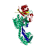
 Basic information
 Components
 Keywords
 Function and homology information
 X-RAY DIFFRACTION /
  Authors
 Citation
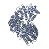
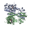
 Structure visualization
 Downloads & links
 Other downloads


 PDBj
PDBj


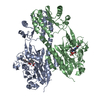
 Assembly

 Mass: 18.015 Da / Num. of mol.: 694 / Source method: isolated from a natural source / Formula: H2O
 Sample preparation
 / Beamline: ID14-1 / Wavelength: 0.934 Å
 Processing