 Movie
Movie Controller

[English] 日本語
 Yorodumi
- PDB-5oq6: Structure of CHK1 12-pt. mutant complex with aminopyrimido-benzod... -
+ Open data

- Basic information
| Entry | Database: PDB / ID: 5oq6 | ||||||
|---|---|---|---|---|---|---|---|
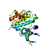
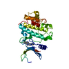
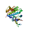
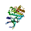
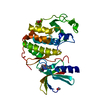
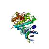
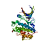
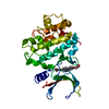
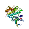
| Title | Structure of CHK1 12-pt. mutant complex with aminopyrimido-benzodiazepinone LRRK2 inhibitor | ||||||
 Components Components | Serine/threonine-protein kinase Chk1 | ||||||
 Keywords Keywords | TRANSFERASE / Parkinson's disease / Leucine-rich repeat kinase 2 / LRRK2 / Checkpoint kinase 1 / CHK1 / mutant / surrogate / kinase inhibitor | ||||||
| Function / homology |  Function and homology information Function and homology informationnegative regulation of mitotic nuclear division / apoptotic process involved in development / negative regulation of G0 to G1 transition / histone H3T11 kinase activity / regulation of mitotic centrosome separation / mitotic G2/M transition checkpoint / inner cell mass cell proliferation / nucleus organization / regulation of double-strand break repair via homologous recombination / peptidyl-threonine phosphorylation ...negative regulation of mitotic nuclear division / apoptotic process involved in development / negative regulation of G0 to G1 transition / histone H3T11 kinase activity / regulation of mitotic centrosome separation / mitotic G2/M transition checkpoint / inner cell mass cell proliferation / nucleus organization / regulation of double-strand break repair via homologous recombination / peptidyl-threonine phosphorylation / mitotic G2 DNA damage checkpoint signaling / negative regulation of gene expression, epigenetic / Transcriptional Regulation by E2F6 / replicative senescence / Presynaptic phase of homologous DNA pairing and strand exchange / Activation of ATR in response to replication stress / Chk1/Chk2(Cds1) mediated inactivation of Cyclin B:Cdk1 complex / signal transduction in response to DNA damage / DNA damage checkpoint signaling / positive regulation of cell cycle / regulation of signal transduction by p53 class mediator / condensed nuclear chromosome / replication fork / TP53 Regulates Transcription of DNA Repair Genes / cellular response to mechanical stimulus / Signaling by SCF-KIT / G2/M DNA damage checkpoint / Ubiquitin-Mediated Degradation of Phosphorylated Cdc25A / G2/M transition of mitotic cell cycle / regulation of cell population proliferation / Processing of DNA double-strand break ends / Regulation of TP53 Activity through Phosphorylation / protein phosphorylation / protein kinase activity / non-specific serine/threonine protein kinase / DNA replication / chromatin remodeling / protein domain specific binding / protein serine kinase activity / DNA repair / protein serine/threonine kinase activity / apoptotic process / centrosome / DNA damage response / chromatin / protein-containing complex / : / nucleoplasm / ATP binding / nucleus / cytosol / cytoplasm Similarity search - Function | ||||||
| Biological species |  Homo sapiens (human) Homo sapiens (human) | ||||||
| Method |  X-RAY DIFFRACTION / SYNCHROTRON / MOLECULAR REPLACEMENT / Resolution: 1.95 Å X-RAY DIFFRACTION / SYNCHROTRON / MOLECULAR REPLACEMENT / Resolution: 1.95 Å | ||||||
 Authors Authors | Dokurno, P. / Williamson, D.S. / Acheson-Dossang, P. / Chen, I. / Murray, J.B. / Shaw, T. / Surgenor, A.E. | ||||||
 Citation Citation | Journal: J. Med. Chem. / Year: 2017 Title: Design of Leucine-Rich Repeat Kinase 2 (LRRK2) Inhibitors Using a Crystallographic Surrogate Derived from Checkpoint Kinase 1 (CHK1). Authors: Williamson, D.S. / Smith, G.P. / Acheson-Dossang, P. / Bedford, S.T. / Chell, V. / Chen, I.J. / Daechsel, J.C.A. / Daniels, Z. / David, L. / Dokurno, P. / Hentzer, M. / Herzig, M.C. / ...Authors: Williamson, D.S. / Smith, G.P. / Acheson-Dossang, P. / Bedford, S.T. / Chell, V. / Chen, I.J. / Daechsel, J.C.A. / Daniels, Z. / David, L. / Dokurno, P. / Hentzer, M. / Herzig, M.C. / Hubbard, R.E. / Moore, J.D. / Murray, J.B. / Newland, S. / Ray, S.C. / Shaw, T. / Surgenor, A.E. / Terry, L. / Thirstrup, K. / Wang, Y. / Christensen, K.V. | ||||||
| History |
|
- Structure visualization
| Structure viewer | Molecule: MolmilJmol/JSmol |
|---|
- Downloads & links
-Download
| PDBx/mmCIF format | 5oq6.cif.gz | 73.7 KB | Display | PDBx/mmCIF format |
|---|---|---|---|---|
| PDB format | pdb5oq6.ent.gz | 52.5 KB | Display | PDB format |
| PDBx/mmJSON format | 5oq6.json.gz | Tree view | PDBx/mmJSON format | |
| Others |  Other downloads Other downloads |
-Validation report
| Arichive directory | https://data.pdbj.org/pub/pdb/validation_reports/oq/5oq6ftp://data.pdbj.org/pub/pdb/validation_reports/oq/5oq6 | HTTPS FTP |
|---|
-Related structure data
| Related structure data | 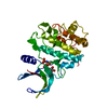 5oopC 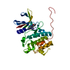 5oorC 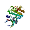 5ootSC 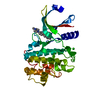 5op2C 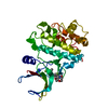 5op4C 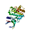 5op5C 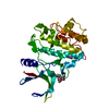 5op7C 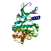 5opbC  5oprC  5opsC 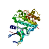 5opuC 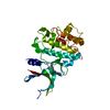 5opvC 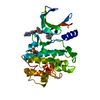 5oq5C 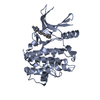 5oq7C 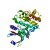 5oq8C S: Starting model for refinement C: citing same article ( |
|---|---|
| Similar structure data |

-Links
 PDBj
PDBj


- Assembly
| Deposited unit | 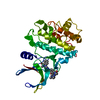
| ||||||||
|---|---|---|---|---|---|---|---|---|---|
| 1 |
| ||||||||
| Unit cell |
|
-Components
| #1: Protein | Mass: 34094.207 Da / Num. of mol.: 1 Mutation: A19S, Y20F, N59L, V68I, L84M, Y86L, C87A, E91S, E134H, S147A, F149Y, G150S Source method: isolated from a genetically manipulated source Source: (gene. exp.) Homo sapiens (human) / Gene: CHEK1, CHK1 / Plasmid: Pfastbac1 / Cell line (production host): SF9 / Production host:   Spodoptera frugiperda (fall armyworm) Spodoptera frugiperda (fall armyworm)References: UniProt: O14757, non-specific serine/threonine protein kinase |
|---|---|
| #2: Chemical | ChemComp-4K4 /   Mass: 570.685 Da / Num. of mol.: 1 / Source method: obtained synthetically / Formula: C31H38N8O3 / Feature type: SUBJECT OF INVESTIGATION Mass: 570.685 Da / Num. of mol.: 1 / Source method: obtained synthetically / Formula: C31H38N8O3 / Feature type: SUBJECT OF INVESTIGATION |
| #3: Chemical | ChemComp-CL /   Mass: 35.453 Da / Num. of mol.: 1 / Source method: obtained synthetically / Formula: Cl Mass: 35.453 Da / Num. of mol.: 1 / Source method: obtained synthetically / Formula: Cl |
| #4: Water | ChemComp-HOH /  Mass: 18.015 Da / Num. of mol.: 165 / Source method: isolated from a natural source / Formula: H2O Mass: 18.015 Da / Num. of mol.: 165 / Source method: isolated from a natural source / Formula: H2O |
-Experimental details
-Experiment
| Experiment | Method: X-RAY DIFFRACTION / Number of used crystals: 1 |
|---|
- Sample preparation
| Crystal | Density Matthews: 2.35 Å3/Da / Density % sol: 47.65 % |
|---|---|
| Crystal grow | Temperature: 293 K / Method: vapor diffusion, sitting drop / pH: 6.5 Details: 7% PEG 8000, 0.1 M MES buffer pH 6.5, 20% ethylene glycol |
-Data collection
| Diffraction | Mean temperature: 100 K | ||||||||||||||||||||||||
|---|---|---|---|---|---|---|---|---|---|---|---|---|---|---|---|---|---|---|---|---|---|---|---|---|---|
| Diffraction source | Source: SYNCHROTRON / Site: Diamond  / Beamline: I03 / Wavelength: 0.9763 Å / Beamline: I03 / Wavelength: 0.9763 Å | ||||||||||||||||||||||||
| Detector | Type: DECTRIS PILATUS3 6M / Detector: PIXEL / Date: Sep 20, 2012 | ||||||||||||||||||||||||
| Radiation | Protocol: SINGLE WAVELENGTH / Monochromatic (M) / Laue (L): M / Scattering type: x-ray | ||||||||||||||||||||||||
| Radiation wavelength | Wavelength: 0.9763 Å / Relative weight: 1 | ||||||||||||||||||||||||
| Reflection | Resolution: 1.91→44.04 Å / Num. obs: 24224 / % possible obs: 98.5 % / Redundancy: 2.9 % / CC1/2: 0.998 / Rmerge(I) obs: 0.051 / Rpim(I) all: 0.036 / Rrim(I) all: 0.063 / Net I/σ(I): 13.7 / Num. measured all: 70334 / Scaling rejects: 0 | ||||||||||||||||||||||||
| Reflection shell | Diffraction-ID: 1
|
- Processing
| Software |
| ||||||||||||||||||||||||||||||||||||||||||||||||||||||||||||
|---|---|---|---|---|---|---|---|---|---|---|---|---|---|---|---|---|---|---|---|---|---|---|---|---|---|---|---|---|---|---|---|---|---|---|---|---|---|---|---|---|---|---|---|---|---|---|---|---|---|---|---|---|---|---|---|---|---|---|---|---|---|
| Refinement | Method to determine structure: MOLECULAR REPLACEMENT Starting model: 5OOT Resolution: 1.95→30 Å / Cor.coef. Fo:Fc: 0.973 / Cor.coef. Fo:Fc free: 0.959 / SU B: 3.97 / SU ML: 0.105 / SU R Cruickshank DPI: 0.1345 / Cross valid method: THROUGHOUT / σ(F): 0 / ESU R: 0.134 / ESU R Free: 0.124 Details: HYDROGENS HAVE BEEN ADDED IN THE RIDING POSITIONS U VALUES : REFINED INDIVIDUALLY
| ||||||||||||||||||||||||||||||||||||||||||||||||||||||||||||
| Solvent computation | Ion probe radii: 0.8 Å / Shrinkage radii: 0.8 Å / VDW probe radii: 1.2 Å | ||||||||||||||||||||||||||||||||||||||||||||||||||||||||||||
| Displacement parameters | Biso max: 117.91 Å2 / Biso mean: 40.027 Å2 / Biso min: 16.98 Å2
| ||||||||||||||||||||||||||||||||||||||||||||||||||||||||||||
| Refinement step | Cycle: final / Resolution: 1.95→30 Å
| ||||||||||||||||||||||||||||||||||||||||||||||||||||||||||||
| Refine LS restraints |
| ||||||||||||||||||||||||||||||||||||||||||||||||||||||||||||
| LS refinement shell | Resolution: 1.95→2.055 Å / Rfactor Rfree error: 0 / Total num. of bins used: 10
|
