 Movie
Movie Controller

[English] 日本語
 Yorodumi
- PDB-5oou: Designed Ankyrin Repeat Protein (DARPin) YTRL-1 selected by direc... -
+ Open data

- Basic information
| Entry | Database: PDB / ID: 5oou | ||||||
|---|---|---|---|---|---|---|---|
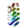
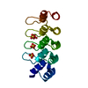
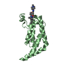
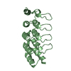
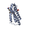
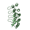
| Title | Designed Ankyrin Repeat Protein (DARPin) YTRL-1 selected by directed evolution against Lysozyme | ||||||
 Components Components | DARPin YTRL-1 | ||||||
 Keywords Keywords | DE NOVO PROTEIN / DARPin / directed evolution / designed protein | ||||||
| Function / homology | Ankyrin repeat-containing domain / Serine Threonine Protein Phosphatase 5, Tetratricopeptide repeat / Alpha Horseshoe / Mainly Alpha Function and homology information Function and homology information | ||||||
| Biological species | synthetic construct (others) | ||||||
| Method |  X-RAY DIFFRACTION / SYNCHROTRON / MOLECULAR REPLACEMENT / Resolution: 2.104 Å X-RAY DIFFRACTION / SYNCHROTRON / MOLECULAR REPLACEMENT / Resolution: 2.104 Å | ||||||
 Authors Authors | Fischer, G. / Hogan, B.J. / Houlihan, G. / Edmond, S. / Huovinen, T.T.K. / Hollfelder, F. / Hyvonen, M. | ||||||
 Citation Citation | Journal: To be published Title: Designed Ankyrin Repeat Protein (DARPin) YTRL-1 selected by directed evolution against Lysozyme Authors: Fischer, G. / Hyvonen, M. | ||||||
| History |
|
- Structure visualization
| Structure viewer | Molecule: MolmilJmol/JSmol |
|---|
- Downloads & links
-Download
| PDBx/mmCIF format | 5oou.cif.gz | 45.1 KB | Display | PDBx/mmCIF format |
|---|---|---|---|---|
| PDB format | pdb5oou.ent.gz | 31.5 KB | Display | PDB format |
| PDBx/mmJSON format | 5oou.json.gz | Tree view | PDBx/mmJSON format | |
| Others |  Other downloads Other downloads |
-Validation report
| Summary document | 5oou_validation.pdf.gz | 412.1 KB | Display | wwPDB validaton report |
|---|---|---|---|---|
| Full document | 5oou_full_validation.pdf.gz | 413.4 KB | Display | |
| Data in XML | 5oou_validation.xml.gz | 8.9 KB | Display | |
| Data in CIF | 5oou_validation.cif.gz | 11.8 KB | Display | |
| Arichive directory | https://data.pdbj.org/pub/pdb/validation_reports/oo/5oouftp://data.pdbj.org/pub/pdb/validation_reports/oo/5oou | HTTPS FTP |
-Related structure data
| Related structure data |  5oosS S: Starting model for refinement |
|---|---|
| Similar structure data |

-Links
 PDBj
PDBj

- Assembly
| Deposited unit | 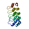
| ||||||||
|---|---|---|---|---|---|---|---|---|---|
| 1 |
| ||||||||
| Unit cell |
|
-Components
| #1: Protein | Mass: 18108.312 Da / Num. of mol.: 1 / Fragment: DARPin / Mutation: YTRL-1 Source method: isolated from a genetically manipulated source Source: (gene. exp.) synthetic construct (others) / Plasmid: pQE30 / Production host:  |
|---|---|
| #2: Water | ChemComp-HOH /  Mass: 18.015 Da / Num. of mol.: 100 / Source method: isolated from a natural source / Formula: H2O Mass: 18.015 Da / Num. of mol.: 100 / Source method: isolated from a natural source / Formula: H2O |
-Experimental details
-Experiment
| Experiment | Method: X-RAY DIFFRACTION / Number of used crystals: 1 |
|---|
- Sample preparation
| Crystal | Density Matthews: 4.1 Å3/Da / Density % sol: 69.96 % |
|---|---|
| Crystal grow | Temperature: 298 K / Method: vapor diffusion / pH: 8.5 / Details: 1.5M AmSO4, 0.1M Tris pH=8.5, 12% glycerol |
-Data collection
| Diffraction | Mean temperature: 100 K | ||||||||||||||||||||||||||||||
|---|---|---|---|---|---|---|---|---|---|---|---|---|---|---|---|---|---|---|---|---|---|---|---|---|---|---|---|---|---|---|---|
| Diffraction source | Source: SYNCHROTRON / Site: Diamond  / Beamline: I03 / Wavelength: 0.97625 Å / Beamline: I03 / Wavelength: 0.97625 Å | ||||||||||||||||||||||||||||||
| Detector | Type: DECTRIS PILATUS 6M / Detector: PIXEL / Date: Mar 11, 2017 | ||||||||||||||||||||||||||||||
| Radiation | Protocol: SINGLE WAVELENGTH / Monochromatic (M) / Laue (L): M / Scattering type: x-ray | ||||||||||||||||||||||||||||||
| Radiation wavelength | Wavelength: 0.97625 Å / Relative weight: 1 | ||||||||||||||||||||||||||||||
| Reflection | Resolution: 2.104→65.557 Å / Num. obs: 18216 / % possible obs: 100 % / Redundancy: 18.9 % / Biso Wilson estimate: 52.04 Å2 / CC1/2: 0.999 / Rmerge(I) obs: 0.1 / Rpim(I) all: 0.024 / Rrim(I) all: 0.103 / Net I/σ(I): 16.5 / Num. measured all: 343934 | ||||||||||||||||||||||||||||||
| Reflection shell | Diffraction-ID: 1
|
- Processing
| Software |
| ||||||||||||||||||||||||||||||||||||||||||
|---|---|---|---|---|---|---|---|---|---|---|---|---|---|---|---|---|---|---|---|---|---|---|---|---|---|---|---|---|---|---|---|---|---|---|---|---|---|---|---|---|---|---|---|
| Refinement | Method to determine structure: MOLECULAR REPLACEMENT Starting model: 5OOS Resolution: 2.104→44.817 Å / SU ML: 0.19 / Cross valid method: THROUGHOUT / σ(F): 1.34 / Phase error: 22.8 / Stereochemistry target values: ML
| ||||||||||||||||||||||||||||||||||||||||||
| Solvent computation | Shrinkage radii: 0.9 Å / VDW probe radii: 1.11 Å / Solvent model: FLAT BULK SOLVENT MODEL | ||||||||||||||||||||||||||||||||||||||||||
| Displacement parameters | Biso max: 163.71 Å2 / Biso mean: 63.404 Å2 / Biso min: 37.3 Å2 | ||||||||||||||||||||||||||||||||||||||||||
| Refinement step | Cycle: final / Resolution: 2.104→44.817 Å
| ||||||||||||||||||||||||||||||||||||||||||
| Refine LS restraints |
| ||||||||||||||||||||||||||||||||||||||||||
| LS refinement shell | Refine-ID: X-RAY DIFFRACTION / Rfactor Rfree error: 0 / Total num. of bins used: 6 / % reflection obs: 100 %
|
