 Movie
Movie Controller

[English] 日本語
 Yorodumi
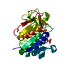
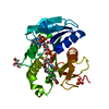
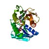
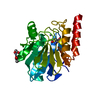
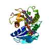
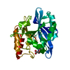
- PDB-5ooh: Human biliverdin IX beta reductase: NADP/Erythrosin extra bluish ... -
+ Open data

- Basic information
| Entry | Database: PDB / ID: 5ooh | ||||||
|---|---|---|---|---|---|---|---|
| Title | Human biliverdin IX beta reductase: NADP/Erythrosin extra bluish ternary complex | ||||||
 Components Components | Flavin reductase (NADPH) | ||||||
 Keywords Keywords | OXIDOREDUCTASE / Biliverdin reductase | ||||||
| Function / homology |  Function and homology information Function and homology informationFMN reductase (NADH) activity / biliverdin reductase [NAD(P)H] activity / flavin reductase (NADPH) / FMN reductase (NADPH) activity / Transferases; Transferring nitrogenous groups; Transferring other nitrogenous groups / Oxidoreductases; Acting on the CH-CH group of donors; With NAD+ or NADP+ as acceptor / megakaryocyte differentiation / riboflavin reductase (NADPH) activity / heme catabolic process / Heme degradation ...FMN reductase (NADH) activity / biliverdin reductase [NAD(P)H] activity / flavin reductase (NADPH) / FMN reductase (NADPH) activity / Transferases; Transferring nitrogenous groups; Transferring other nitrogenous groups / Oxidoreductases; Acting on the CH-CH group of donors; With NAD+ or NADP+ as acceptor / megakaryocyte differentiation / riboflavin reductase (NADPH) activity / heme catabolic process / Heme degradation / peptidyl-cysteine S-nitrosylase activity / negative regulation of insulin receptor signaling pathway / Cytoprotection by HMOX1 / extracellular exosome / nucleoplasm / plasma membrane / cytoplasm / cytosol Similarity search - Function | ||||||
| Biological species |  Homo sapiens (human) Homo sapiens (human) | ||||||
| Method |  X-RAY DIFFRACTION / SYNCHROTRON / MOLECULAR REPLACEMENT / Resolution: 1.2 Å X-RAY DIFFRACTION / SYNCHROTRON / MOLECULAR REPLACEMENT / Resolution: 1.2 Å | ||||||
 Authors Authors | Manso, J.A. / Pereira, P.J.B. | ||||||
 Citation Citation | Journal: J. Biol. Chem. / Year: 2018 Title: In silicoand crystallographic studies identify key structural features of biliverdin IX beta reductase inhibitors having nanomolar potency. Authors: Nesbitt, N.M. / Zheng, X. / Li, Z. / Manso, J.A. / Yen, W.Y. / Malone, L.E. / Ripoll-Rozada, J. / Pereira, P.J.B. / Mantle, T.J. / Wang, J. / Bahou, W.F. | ||||||
| History |
|
- Structure visualization
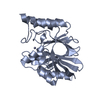
| Structure viewer | Molecule: MolmilJmol/JSmol |
|---|
- Downloads & links
-Download
| PDBx/mmCIF format | 5ooh.cif.gz | 146.2 KB | Display | PDBx/mmCIF format |
|---|---|---|---|---|
| PDB format | pdb5ooh.ent.gz | 116.2 KB | Display | PDB format |
| PDBx/mmJSON format | 5ooh.json.gz | Tree view | PDBx/mmJSON format | |
| Others |  Other downloads Other downloads |
-Validation report
| Arichive directory | https://data.pdbj.org/pub/pdb/validation_reports/oo/5oohftp://data.pdbj.org/pub/pdb/validation_reports/oo/5ooh | HTTPS FTP |
|---|
-Related structure data
| Related structure data |  5oogC  1hdoS S: Starting model for refinement C: citing same article ( |
|---|---|
| Similar structure data | |
| Experimental dataset #1 | Data reference: 10.15785/SBGRID/486 / Data set type: diffraction image data |
| Experimental dataset #2 | Data reference: 10.15785/SBGRID/487 / Data set type: diffraction image data |


-Links
 PDBj
PDBj

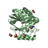
- Assembly
| Deposited unit | 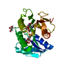
| ||||||||
|---|---|---|---|---|---|---|---|---|---|
| 1 |
| ||||||||
| Unit cell |
|
-Components
| #1: Protein | Mass: 22148.350 Da / Num. of mol.: 1 Source method: isolated from a genetically manipulated source Source: (gene. exp.) Homo sapiens (human) / Gene: BLVRB, FLR / Production host:  References: UniProt: P30043, flavin reductase (NADPH), biliverdin reductase |
|---|---|
| #2: Chemical | ChemComp-NAP /   Mass: 743.405 Da / Num. of mol.: 1 Mass: 743.405 Da / Num. of mol.: 1Source method: isolated from a genetically manipulated source Formula: C21H28N7O17P3 |
| #3: Chemical | ChemComp-GOL /   Mass: 92.094 Da / Num. of mol.: 1 / Source method: obtained synthetically / Formula: C3H8O3 Mass: 92.094 Da / Num. of mol.: 1 / Source method: obtained synthetically / Formula: C3H8O3 |
| #4: Chemical | ChemComp-9ZZ / 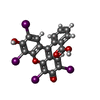  Mass: 835.892 Da / Num. of mol.: 1 / Source method: obtained synthetically / Formula: C20H8I4O5 / Feature type: SUBJECT OF INVESTIGATION Mass: 835.892 Da / Num. of mol.: 1 / Source method: obtained synthetically / Formula: C20H8I4O5 / Feature type: SUBJECT OF INVESTIGATION |
| #5: Water | ChemComp-HOH /  Mass: 18.015 Da / Num. of mol.: 319 / Source method: isolated from a natural source / Formula: H2O Mass: 18.015 Da / Num. of mol.: 319 / Source method: isolated from a natural source / Formula: H2O |
-Experimental details
-Experiment
| Experiment | Method: X-RAY DIFFRACTION / Number of used crystals: 1 |
|---|
- Sample preparation
| Crystal | Density Matthews: 2.4 Å3/Da / Density % sol: 48.76 % |
|---|---|
| Crystal grow | Temperature: 293 K / Method: vapor diffusion, hanging drop Details: 30% PEG 3350, 0.2 M AMMONIUM SULFATE, 0.1 M SODIUM CACODYLATE PH 6.5. NADP ADDED TO A FINAL CONCENTRATION OF 2.5 MM |
-Data collection
| Diffraction | Mean temperature: 100 K |
|---|---|
| Diffraction source | Source: SYNCHROTRON / Site: ESRF  / Beamline: ID23-1 / Wavelength: 0.9763 Å / Beamline: ID23-1 / Wavelength: 0.9763 Å |
| Detector | Type: ADSC QUANTUM 315r / Detector: CCD / Date: Apr 14, 2010 |
| Radiation | Protocol: SINGLE WAVELENGTH / Monochromatic (M) / Laue (L): M / Scattering type: x-ray |
| Radiation wavelength | Wavelength: 0.9763 Å / Relative weight: 1 |
| Reflection | Resolution: 1.2→40.3 Å / Num. obs: 65182 / % possible obs: 96.6 % / Redundancy: 10.1 % / Biso Wilson estimate: 3.9 Å2 / CC1/2: 0.999 / Rmerge(I) obs: 0.106 / Rpim(I) all: 0.034 / Rrim(I) all: 0.111 / Net I/σ(I): 15.3 |
| Reflection shell | Resolution: 1.2→1.22 Å / Redundancy: 8.3 % / Rmerge(I) obs: 0.838 / Mean I/σ(I) obs: 3.1 / Num. unique obs: 3061 / CC1/2: 0.808 / Rpim(I) all: 0.308 / Rrim(I) all: 0.893 / % possible all: 92.4 |
- Processing
| Software |
| |||||||||||||||||||||||||||||||||||||||||||||||||||||||||||||||||||||||||||||||||||||||||||||||||||||||||||||||||||||||||||||||||||||||||||||||||||||||||||||||||||||||||||||||||||||||||||||||||||||||||||||||||||||||||
|---|---|---|---|---|---|---|---|---|---|---|---|---|---|---|---|---|---|---|---|---|---|---|---|---|---|---|---|---|---|---|---|---|---|---|---|---|---|---|---|---|---|---|---|---|---|---|---|---|---|---|---|---|---|---|---|---|---|---|---|---|---|---|---|---|---|---|---|---|---|---|---|---|---|---|---|---|---|---|---|---|---|---|---|---|---|---|---|---|---|---|---|---|---|---|---|---|---|---|---|---|---|---|---|---|---|---|---|---|---|---|---|---|---|---|---|---|---|---|---|---|---|---|---|---|---|---|---|---|---|---|---|---|---|---|---|---|---|---|---|---|---|---|---|---|---|---|---|---|---|---|---|---|---|---|---|---|---|---|---|---|---|---|---|---|---|---|---|---|---|---|---|---|---|---|---|---|---|---|---|---|---|---|---|---|---|---|---|---|---|---|---|---|---|---|---|---|---|---|---|---|---|---|---|---|---|---|---|---|---|---|---|---|---|---|---|---|---|---|
| Refinement | Method to determine structure: MOLECULAR REPLACEMENT Starting model: 1HDO Resolution: 1.2→32.194 Å / SU ML: 0.1 / Cross valid method: FREE R-VALUE / σ(F): 0.01 / Phase error: 12.45
| |||||||||||||||||||||||||||||||||||||||||||||||||||||||||||||||||||||||||||||||||||||||||||||||||||||||||||||||||||||||||||||||||||||||||||||||||||||||||||||||||||||||||||||||||||||||||||||||||||||||||||||||||||||||||
| Solvent computation | Shrinkage radii: 0.9 Å / VDW probe radii: 1.11 Å | |||||||||||||||||||||||||||||||||||||||||||||||||||||||||||||||||||||||||||||||||||||||||||||||||||||||||||||||||||||||||||||||||||||||||||||||||||||||||||||||||||||||||||||||||||||||||||||||||||||||||||||||||||||||||
| Refinement step | Cycle: LAST / Resolution: 1.2→32.194 Å
| |||||||||||||||||||||||||||||||||||||||||||||||||||||||||||||||||||||||||||||||||||||||||||||||||||||||||||||||||||||||||||||||||||||||||||||||||||||||||||||||||||||||||||||||||||||||||||||||||||||||||||||||||||||||||
| Refine LS restraints |
| |||||||||||||||||||||||||||||||||||||||||||||||||||||||||||||||||||||||||||||||||||||||||||||||||||||||||||||||||||||||||||||||||||||||||||||||||||||||||||||||||||||||||||||||||||||||||||||||||||||||||||||||||||||||||
| LS refinement shell |
|
