Mass: 18.015 Da / Num. of mol.: 37 / Source method: isolated from a natural source / Formula: H2O
Has protein modification
Y
-
Experimental details
-
Experiment
Experiment
Method: X-RAY DIFFRACTION / Number of used crystals: 1
-
Sample preparation
Crystal
Density Matthews: 2.49 Å3/Da / Density % sol: 50.6 %
Crystal grow
Temperature: 293 K / Method: vapor diffusion, hanging drop / pH: 6.8 Details: 100 mM MES, 5 mM MgCl2, 5 mM CaCl2, 50 mM CdCl2, 50 mM NiCl2 and 12% (w/v) PEG 3350
Method to determine structure: SAD / Resolution: 1.45→12 Å / Cor.coef. Fo:Fc: 0.964 / Cor.coef. Fo:Fc free: 0.957 / SU R Cruickshank DPI: 0.069 / Cross valid method: THROUGHOUT / σ(F): 0 / SU R Blow DPI: 0.071 / SU Rfree Blow DPI: 0.063 / SU Rfree Cruickshank DPI: 0.062
Rfactor
Num. reflection
% reflection
Selection details
Rfree
0.191
362
5 %
RANDOM
Rwork
0.19
-
-
-
obs
0.19
7238
100 %
-
Displacement parameters
Biso mean: 34.98 Å2
Baniso -1
Baniso -2
Baniso -3
1-
-0.6515 Å2
0 Å2
0 Å2
2-
-
-0.6515 Å2
0 Å2
3-
-
-
1.3029 Å2
Refine analyze
Luzzati coordinate error obs: 0.19 Å
Refinement step
Cycle: 1 / Resolution: 1.45→12 Å
Protein
Nucleic acid
Ligand
Solvent
Total
Num. atoms
293
0
3
37
333
Refine LS restraints
Refine-ID
Type
Dev ideal
Number
Restraint function
Weight
X-RAY DIFFRACTION
t_bond_d
0.01
341
HARMONIC
2
X-RAY DIFFRACTION
t_angle_deg
1.13
469
HARMONIC
2
X-RAY DIFFRACTION
t_dihedral_angle_d
122
SINUSOIDAL
2
X-RAY DIFFRACTION
t_incorr_chiral_ct
X-RAY DIFFRACTION
t_pseud_angle
X-RAY DIFFRACTION
t_trig_c_planes
11
HARMONIC
2
X-RAY DIFFRACTION
t_gen_planes
47
HARMONIC
5
X-RAY DIFFRACTION
t_it
341
HARMONIC
20
X-RAY DIFFRACTION
t_nbd
0
SEMIHARMONIC
5
X-RAY DIFFRACTION
t_omega_torsion
4.86
X-RAY DIFFRACTION
t_other_torsion
18.96
X-RAY DIFFRACTION
t_improper_torsion
X-RAY DIFFRACTION
t_chiral_improper_torsion
38
SEMIHARMONIC
5
X-RAY DIFFRACTION
t_sum_occupancies
X-RAY DIFFRACTION
t_utility_distance
4
HARMONIC
1
X-RAY DIFFRACTION
t_utility_angle
X-RAY DIFFRACTION
t_utility_torsion
X-RAY DIFFRACTION
t_ideal_dist_contact
437
SEMIHARMONIC
4
LS refinement shell
Resolution: 1.45→1.62 Å / Total num. of bins used: 5
Rfactor
Num. reflection
% reflection
Rfree
0.262
103
5.01 %
Rwork
0.2301
1952
-
all
0.2316
2055
-
obs
-
-
100 %
Refinement TLS params.
Method: refined / Origin x: 4.0763 Å / Origin y: 10.6025 Å / Origin z: 15.9414 Å
11
12
13
21
22
23
31
32
33
T
0.0261 Å2
-0.014 Å2
-0.0812 Å2
-
-0.0111 Å2
0.0491 Å2
-
-
0.0884 Å2
L
2.9403 °2
1.7049 °2
-0.7095 °2
-
4.6367 °2
1.08 °2
-
-
2.378 °2
S
-0.205 Å °
-0.0156 Å °
0.4939 Å °
-0.3065 Å °
-0.0174 Å °
-0.0786 Å °
-0.1273 Å °
-0.0444 Å °
0.2224 Å °
Refinement TLS group
Selection details: { *|* }
+
About Yorodumi
-
News
-
Feb 9, 2022. New format data for meta-information of EMDB entries
New format data for meta-information of EMDB entries
Version 3 of the EMDB header file is now the official format.
The previous official version 1.9 will be removed from the archive.
In the structure databanks used in Yorodumi, some data are registered as the other names, "COVID-19 virus" and "2019-nCoV". Here are the details of the virus and the list of structure data.
Jan 31, 2019. EMDB accession codes are about to change! (news from PDBe EMDB page)
EMDB accession codes are about to change! (news from PDBe EMDB page)
The allocation of 4 digits for EMDB accession codes will soon come to an end. Whilst these codes will remain in use, new EMDB accession codes will include an additional digit and will expand incrementally as the available range of codes is exhausted. The current 4-digit format prefixed with “EMD-” (i.e. EMD-XXXX) will advance to a 5-digit format (i.e. EMD-XXXXX), and so on. It is currently estimated that the 4-digit codes will be depleted around Spring 2019, at which point the 5-digit format will come into force.
The EM Navigator/Yorodumi systems omit the EMD- prefix.
Related info.:Q: What is EMD? / ID/Accession-code notation in Yorodumi/EM Navigator
Yorodumi is a browser for structure data from EMDB, PDB, SASBDB, etc.
This page is also the successor to EM Navigator detail page, and also detail information page/front-end page for Omokage search.
The word "yorodu" (or yorozu) is an old Japanese word meaning "ten thousand". "mi" (miru) is to see.
Related info.:EMDB / PDB / SASBDB / Comparison of 3 databanks / Yorodumi Search / Aug 31, 2016. New EM Navigator & Yorodumi / Yorodumi Papers / Jmol/JSmol / Function and homology information / Changes in new EM Navigator and Yorodumi
 Movie
Movie Controller

 Yorodumi
 Open data
 Basic information
 Components
 Keywords
 Function and homology information
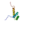
 Gymnema sylvestre (plant)
 X-RAY DIFFRACTION /
  Authors
 Citation
 Structure visualization
 Downloads & links
 Other downloads

 PDBj
PDBj
 Assembly

 Komagataella phaffii GS115 (fungus) / References: UniProt: P25810
 Mass: 58.693 Da / Num. of mol.: 3 / Source method: obtained synthetically / Formula: Ni
 Mass: 18.015 Da / Num. of mol.: 37 / Source method: isolated from a natural source / Formula: H2O
 Sample preparation
 / Beamline: PROXIMA 1 / Wavelength: 1.0332 Å
 Processing