 Movie
Movie Controller

[English] 日本語
 Yorodumi
- PDB-5olj: Crystal structure of Porphyromonas gingivalis dipeptidyl peptidase 4 -
+ Open data

- Basic information
| Entry | Database: PDB / ID: 5olj | |||||||||||||||
|---|---|---|---|---|---|---|---|---|---|---|---|---|---|---|---|---|
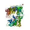
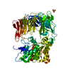
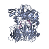
| Title | Crystal structure of Porphyromonas gingivalis dipeptidyl peptidase 4 | |||||||||||||||
 Components Components | Dipeptidyl peptidase IV | |||||||||||||||
 Keywords Keywords | HYDROLASE / dipeptidyl peptidase 4 / peptidase inhibitor / Porphyromonas gingivalis / biofilm / dipeptidyl peptidase 9 | |||||||||||||||
| Function / homology |  Function and homology information Function and homology information | |||||||||||||||
| Biological species |  Porphyromonas gingivalis (bacteria) Porphyromonas gingivalis (bacteria) | |||||||||||||||
| Method |  X-RAY DIFFRACTION / MOLECULAR REPLACEMENT / Resolution: 2.2 Å X-RAY DIFFRACTION / MOLECULAR REPLACEMENT / Resolution: 2.2 Å | |||||||||||||||
 Authors Authors | Fulop, V. | |||||||||||||||
| Funding support |  Belgium, 4items Belgium, 4items
| |||||||||||||||
 Citation Citation | Journal: Eur J Med Chem / Year: 2017 Title: Crystal structure of Porphyromonas gingivalis dipeptidyl peptidase 4 and structure-activity relationships based on inhibitor profiling. Authors: Rea, D. / Van Elzen, R. / De Winter, H. / Van Goethem, S. / Landuyt, B. / Luyten, W. / Schoofs, L. / Van Der Veken, P. / Augustyns, K. / De Meester, I. / Fulop, V. / Lambeir, A.M. #1: Journal: Acta Crystallogr. D Biol. Crystallogr. / Year: 2004 Title: Expression, purification and preliminary crystallographic analysis of dipeptidyl peptidase IV from Porphyromonas gingivalis. Authors: Rea, D. / Lambeir, A.M. / Kumagai, Y. / De Meester, I. / Scharpe, S. / Fulop, V. | |||||||||||||||
| History |
|
- Structure visualization
| Structure viewer | Molecule: MolmilJmol/JSmol |
|---|
- Downloads & links
-Download
| PDBx/mmCIF format | 5olj.cif.gz | 271.6 KB | Display | PDBx/mmCIF format |
|---|---|---|---|---|
| PDB format | pdb5olj.ent.gz | 215.9 KB | Display | PDB format |
| PDBx/mmJSON format | 5olj.json.gz | Tree view | PDBx/mmJSON format | |
| Others |  Other downloads Other downloads |
-Validation report
| Arichive directory | https://data.pdbj.org/pub/pdb/validation_reports/ol/5oljftp://data.pdbj.org/pub/pdb/validation_reports/ol/5olj | HTTPS FTP |
|---|
-Related structure data
| Related structure data |  5kbyS S: Starting model for refinement |
|---|---|
| Similar structure data |


-Links
 PDBj
PDBj
- Assembly
| Deposited unit | 
| |||||||||
|---|---|---|---|---|---|---|---|---|---|---|
| 1 |
| |||||||||
| Unit cell |
| |||||||||
| Components on special symmetry positions |
|
-Components
| #1: Protein | Mass: 82087.719 Da / Num. of mol.: 1 Source method: isolated from a genetically manipulated source Source: (gene. exp.) Porphyromonas gingivalis (bacteria) / Gene: dppIV, PGN_1469 / Production host: Porphyromonas gingivalis (bacteria) / References: UniProt: B2RKU3 |
|---|---|
| #2: Chemical | ChemComp-GOL /   Mass: 92.094 Da / Num. of mol.: 1 / Source method: isolated from a natural source / Formula: C3H8O3 Mass: 92.094 Da / Num. of mol.: 1 / Source method: isolated from a natural source / Formula: C3H8O3 |
| #3: Water | ChemComp-HOH /  Mass: 18.015 Da / Num. of mol.: 398 / Source method: isolated from a natural source / Formula: H2O Mass: 18.015 Da / Num. of mol.: 398 / Source method: isolated from a natural source / Formula: H2O |
-Experimental details
-Experiment
| Experiment | Method: X-RAY DIFFRACTION / Number of used crystals: 1 |
|---|
- Sample preparation
| Crystal | Density Matthews: 2.86 Å3/Da / Density % sol: 57 % |
|---|---|
| Crystal grow | Temperature: 290 K / Method: vapor diffusion Details: 10% (w/v) PEG 8000, 20% (v/v) ethylene glycol, 0.02 M sodium L-glutamate, 0.02 M DL-alanine, 0.02 M glycine, 0.02 M DL-lysine, 0.02 M DLserine, and 0.1 M BICINE/TRIS pH 8.5 PH range: 8.5 |
-Data collection
| Diffraction | Mean temperature: 100 K |
|---|---|
| Diffraction source | Source: SEALED TUBE / Type: Xenocs GeniX 3D Cu HF / Wavelength: 1.5418 Å |
| Detector | Type: MARRESEARCH / Detector: IMAGE PLATE / Date: Jul 9, 2011 |
| Radiation | Protocol: SINGLE WAVELENGTH / Monochromatic (M) / Laue (L): M / Scattering type: x-ray |
| Radiation wavelength | Wavelength: 1.5418 Å / Relative weight: 1 |
| Reflection | Resolution: 2.2→38 Å / Num. obs: 48811 / % possible obs: 99.7 % / Redundancy: 9.3 % / Net I/σ(I): 10.8 |
- Processing
| Software | Name: REFMAC / Version: 5.7.0029 / Classification: refinement | ||||||||||||||||||||||||||||||||||||||||||||||||||||||||||||||||||||||||||||||||||||||||||||||||||||||||||||||||||||||||||||||||||||||||||||||||||||||||||||||||||||||||||||||||||||||
|---|---|---|---|---|---|---|---|---|---|---|---|---|---|---|---|---|---|---|---|---|---|---|---|---|---|---|---|---|---|---|---|---|---|---|---|---|---|---|---|---|---|---|---|---|---|---|---|---|---|---|---|---|---|---|---|---|---|---|---|---|---|---|---|---|---|---|---|---|---|---|---|---|---|---|---|---|---|---|---|---|---|---|---|---|---|---|---|---|---|---|---|---|---|---|---|---|---|---|---|---|---|---|---|---|---|---|---|---|---|---|---|---|---|---|---|---|---|---|---|---|---|---|---|---|---|---|---|---|---|---|---|---|---|---|---|---|---|---|---|---|---|---|---|---|---|---|---|---|---|---|---|---|---|---|---|---|---|---|---|---|---|---|---|---|---|---|---|---|---|---|---|---|---|---|---|---|---|---|---|---|---|---|---|
| Refinement | Method to determine structure: MOLECULAR REPLACEMENT Starting model: 5KBY Resolution: 2.2→37.87 Å / Cor.coef. Fo:Fc: 0.839 / Cor.coef. Fo:Fc free: 0.81 / SU B: 9.784 / SU ML: 0.126 / Cross valid method: THROUGHOUT / ESU R: 0.258 / ESU R Free: 0.204 / Stereochemistry target values: MAXIMUM LIKELIHOOD / Details: HYDROGENS HAVE BEEN ADDED IN THE RIDING POSITIONS
| ||||||||||||||||||||||||||||||||||||||||||||||||||||||||||||||||||||||||||||||||||||||||||||||||||||||||||||||||||||||||||||||||||||||||||||||||||||||||||||||||||||||||||||||||||||||
| Solvent computation | Ion probe radii: 0.8 Å / Shrinkage radii: 0.8 Å / VDW probe radii: 1.2 Å / Solvent model: MASK | ||||||||||||||||||||||||||||||||||||||||||||||||||||||||||||||||||||||||||||||||||||||||||||||||||||||||||||||||||||||||||||||||||||||||||||||||||||||||||||||||||||||||||||||||||||||
| Displacement parameters | Biso mean: 4.429 Å2
| ||||||||||||||||||||||||||||||||||||||||||||||||||||||||||||||||||||||||||||||||||||||||||||||||||||||||||||||||||||||||||||||||||||||||||||||||||||||||||||||||||||||||||||||||||||||
| Refinement step | Cycle: 1 / Resolution: 2.2→37.87 Å
| ||||||||||||||||||||||||||||||||||||||||||||||||||||||||||||||||||||||||||||||||||||||||||||||||||||||||||||||||||||||||||||||||||||||||||||||||||||||||||||||||||||||||||||||||||||||
| Refine LS restraints |
|