Mass: 18.015 Da / Num. of mol.: 516 / Source method: isolated from a natural source / Formula: H2O
-
Experimental details
-
Experiment
Experiment
Method: X-RAY DIFFRACTION / Number of used crystals: 1
-
Sample preparation
Crystal
Density Matthews: 2.93 Å3/Da / Density % sol: 58.04 % Description: Transparent cube shape of about 100x100 micrometer.
Crystal grow
Temperature: 281.15 K / Method: vapor diffusion, sitting drop / pH: 7 Details: 1-ml mixture containing 120 mM potassium phosphate pH 6.0, 1 mM EDTA, 26 uM (1 mg/ml) [Fe]-hydrogenase and 26 uM methylene-H4MPT was incubated in 5-ml amber vials with rubber stoppers under ...Details: 1-ml mixture containing 120 mM potassium phosphate pH 6.0, 1 mM EDTA, 26 uM (1 mg/ml) [Fe]-hydrogenase and 26 uM methylene-H4MPT was incubated in 5-ml amber vials with rubber stoppers under a gas mixture of N2/O2/H2 (70%/20%/10%) at 40 degree Celsius for 1 h. The inactivated enzyme was concentrated to 25 mg/ml using a 30 kDa centrifugal filter (Millipore). 0.7-ul of 25 mg/ml O2-inactivated [Fe]-hydrogenase was mixed with 0.7 ul of reservoir solution of 2 M (NH4)2SO4, 100 mM Tris/HCl, pH 7.0, and 200 mM LiSO4. The best diffracting crystal was obtained in one month. Prior to freezing the crystal was soaked for 3 seconds in 2 M (NH4)2SO4, 100 mM Tris/HCl, pH 7.0, 200 mM LiSO4 and 30% glycerol v/v.
In the structure databanks used in Yorodumi, some data are registered as the other names, "COVID-19 virus" and "2019-nCoV". Here are the details of the virus and the list of structure data.
Jan 31, 2019. EMDB accession codes are about to change! (news from PDBe EMDB page)
EMDB accession codes are about to change! (news from PDBe EMDB page)
The allocation of 4 digits for EMDB accession codes will soon come to an end. Whilst these codes will remain in use, new EMDB accession codes will include an additional digit and will expand incrementally as the available range of codes is exhausted. The current 4-digit format prefixed with “EMD-” (i.e. EMD-XXXX) will advance to a 5-digit format (i.e. EMD-XXXXX), and so on. It is currently estimated that the 4-digit codes will be depleted around Spring 2019, at which point the 5-digit format will come into force.
The EM Navigator/Yorodumi systems omit the EMD- prefix.
Related info.:Q: What is EMD? / ID/Accession-code notation in Yorodumi/EM Navigator
Yorodumi is a browser for structure data from EMDB, PDB, SASBDB, etc.
This page is also the successor to EM Navigator detail page, and also detail information page/front-end page for Omokage search.
The word "yorodu" (or yorozu) is an old Japanese word meaning "ten thousand". "mi" (miru) is to see.
Related info.:EMDB / PDB / SASBDB / Comparison of 3 databanks / Yorodumi Search / Aug 31, 2016. New EM Navigator & Yorodumi / Yorodumi Papers / Jmol/JSmol / Function and homology information / Changes in new EM Navigator and Yorodumi
 Movie
Movie Controller

 Yorodumi
 Open data
 Basic information
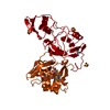
 Components
 Keywords
 Function and homology information
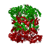
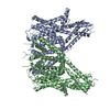
 Methanothermobacter marburgensis str. Marburg (archaea)
 X-RAY DIFFRACTION /
  Authors
 Citation
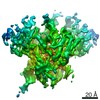
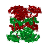
 Structure visualization
 Downloads & links
 Other downloads
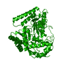

 PDBj
PDBj


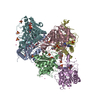
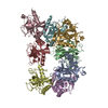
 Assembly
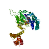

 Mass: 55.845 Da / Num. of mol.: 3 / Source method: obtained synthetically / Formula: Fe
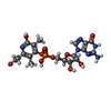
 Mass: 542.393 Da / Num. of mol.: 1 / Source method: obtained synthetically / Formula: C19H23N6O11P
 Mass: 92.094 Da / Num. of mol.: 1 / Source method: obtained synthetically / Formula: C3H8O3
 Mass: 18.015 Da / Num. of mol.: 516 / Source method: isolated from a natural source / Formula: H2O
 Sample preparation
 / Beamline: X10SA / Wavelength: 1.00005 Å
 Processing