 Movie
Movie Controller

[English] 日本語
 Yorodumi
- PDB-5oiz: Penicillin-Binding Protein 2X (PBP2X) from Streptococcus pneumoni... -
+ Open data

- Basic information
| Entry | Database: PDB / ID: 5oiz | ||||||
|---|---|---|---|---|---|---|---|
| Title | Penicillin-Binding Protein 2X (PBP2X) from Streptococcus pneumoniae in complex with oxacillin | ||||||
 Components Components | Penicillin-binding protein 2X | ||||||
 Keywords Keywords | ANTIBIOTIC / Penicillin / b-lactam / cell-wall / transpeptidase | ||||||
| Function / homology |  Function and homology information Function and homology informationpenicillin binding / peptidoglycan biosynthetic process / cell wall organization / regulation of cell shape / cell division / response to antibiotic / plasma membrane Similarity search - Function | ||||||
| Biological species |   Streptococcus pneumoniae (bacteria) Streptococcus pneumoniae (bacteria) | ||||||
| Method |  X-RAY DIFFRACTION / SYNCHROTRON / MOLECULAR REPLACEMENT / Resolution: 2.7 Å X-RAY DIFFRACTION / SYNCHROTRON / MOLECULAR REPLACEMENT / Resolution: 2.7 Å | ||||||
 Authors Authors | Bernardo-Garcia, N. / Hermoso, J.A. | ||||||
 Citation Citation | Journal: ACS Chem. Biol. / Year: 2018 Title: Allostery, Recognition of Nascent Peptidoglycan, and Cross-linking of the Cell Wall by the Essential Penicillin-Binding Protein 2x of Streptococcus pneumoniae. Authors: Bernardo-Garcia, N. / Mahasenan, K.V. / Batuecas, M.T. / Lee, M. / Hesek, D. / Petrackova, D. / Doubravova, L. / Branny, P. / Mobashery, S. / Hermoso, J.A. | ||||||
| History |
|
- Structure visualization
| Structure viewer | Molecule: MolmilJmol/JSmol |
|---|
- Downloads & links
-Download
| PDBx/mmCIF format | 5oiz.cif.gz | 260.8 KB | Display | PDBx/mmCIF format |
|---|---|---|---|---|
| PDB format | pdb5oiz.ent.gz | 208 KB | Display | PDB format |
| PDBx/mmJSON format | 5oiz.json.gz | Tree view | PDBx/mmJSON format | |
| Others |  Other downloads Other downloads |
-Validation report
| Arichive directory | https://data.pdbj.org/pub/pdb/validation_reports/oi/5oizftp://data.pdbj.org/pub/pdb/validation_reports/oi/5oiz | HTTPS FTP |
|---|
-Related structure data
| Related structure data | 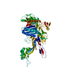 5oauC 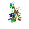 5oj0C 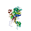 5oj1C 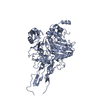 1k25S S: Starting model for refinement C: citing same article ( |
|---|---|
| Similar structure data |


-Links
 PDBj
PDBj- Assembly
| Deposited unit | 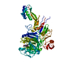
| ||||||||
|---|---|---|---|---|---|---|---|---|---|
| 1 |
| ||||||||
| Unit cell |
|
-Components
| #1: Protein | Mass: 76807.969 Da / Num. of mol.: 1 Source method: isolated from a genetically manipulated source Source: (gene. exp.) Streptococcus pneumoniae (strain ATCC BAA-255 / R6) (bacteria)Gene: pbpX, spr0304 / Production host: |
|---|---|
| #2: Chemical | ChemComp-1S6 / (  Mass: 403.452 Da / Num. of mol.: 1 / Source method: obtained synthetically / Formula: C19H21N3O5S / Comment: antibiotic*YM Mass: 403.452 Da / Num. of mol.: 1 / Source method: obtained synthetically / Formula: C19H21N3O5S / Comment: antibiotic*YM |
| #3: Water | ChemComp-HOH /  Mass: 18.015 Da / Num. of mol.: 100 / Source method: isolated from a natural source / Formula: H2O Mass: 18.015 Da / Num. of mol.: 100 / Source method: isolated from a natural source / Formula: H2O |
| Has protein modification | Y |
-Experimental details
-Experiment
| Experiment | Method: X-RAY DIFFRACTION / Number of used crystals: 1 |
|---|
- Sample preparation
| Crystal | Density Matthews: 3.56 Å3/Da / Density % sol: 65.45 % |
|---|---|
| Crystal grow | Temperature: 277 K / Method: vapor diffusion, sitting drop / pH: 4.5 / Details: 2.3-3.0 M NaCl, 0.1 M sodium acetate pH 4.5 |
-Data collection
| Diffraction | Mean temperature: 100 K |
|---|---|
| Diffraction source | Source: SYNCHROTRON / Site: ALBA  / Beamline: XALOC / Wavelength: 0.97924 Å / Beamline: XALOC / Wavelength: 0.97924 Å |
| Detector | Type: DECTRIS PILATUS 6M / Detector: PIXEL / Date: Sep 17, 2015 |
| Radiation | Protocol: SINGLE WAVELENGTH / Monochromatic (M) / Laue (L): M / Scattering type: x-ray |
| Radiation wavelength | Wavelength: 0.97924 Å / Relative weight: 1 |
| Reflection | Resolution: 2.7→86.79 Å / Num. obs: 30875 / % possible obs: 99.7 % / Redundancy: 3.8 % / CC1/2: 0.99 / Rmerge(I) obs: 0.137 / Rpim(I) all: 0.078 / Net I/σ(I): 8.5 |
| Reflection shell | Resolution: 2.7→2.83 Å / Redundancy: 3.8 % / Rmerge(I) obs: 1.14 / Mean I/σ(I) obs: 1.2 / Num. unique obs: 4451 / CC1/2: 0.58 / Rpim(I) all: 0.67 / % possible all: 99.9 |
- Processing
| Software |
| ||||||||||||||||||||||||||||||||||||||||||||||||||||||||||||||||||||||||||||||||||||||||||||||||||||||||||||||||||||||||||||||||||||||||||||||||||||||||||||||||||||||||||||||||||||||
|---|---|---|---|---|---|---|---|---|---|---|---|---|---|---|---|---|---|---|---|---|---|---|---|---|---|---|---|---|---|---|---|---|---|---|---|---|---|---|---|---|---|---|---|---|---|---|---|---|---|---|---|---|---|---|---|---|---|---|---|---|---|---|---|---|---|---|---|---|---|---|---|---|---|---|---|---|---|---|---|---|---|---|---|---|---|---|---|---|---|---|---|---|---|---|---|---|---|---|---|---|---|---|---|---|---|---|---|---|---|---|---|---|---|---|---|---|---|---|---|---|---|---|---|---|---|---|---|---|---|---|---|---|---|---|---|---|---|---|---|---|---|---|---|---|---|---|---|---|---|---|---|---|---|---|---|---|---|---|---|---|---|---|---|---|---|---|---|---|---|---|---|---|---|---|---|---|---|---|---|---|---|---|---|
| Refinement | Method to determine structure: MOLECULAR REPLACEMENT Starting model: 1K25 Resolution: 2.7→48.54 Å / Cor.coef. Fo:Fc: 0.945 / Cor.coef. Fo:Fc free: 0.908 / SU B: 10.963 / SU ML: 0.117 / Cross valid method: THROUGHOUT / ESU R: 0.095 / ESU R Free: 0.068 / Stereochemistry target values: MAXIMUM LIKELIHOOD / Details: HYDROGENS HAVE BEEN ADDED IN THE RIDING POSITIONS
| ||||||||||||||||||||||||||||||||||||||||||||||||||||||||||||||||||||||||||||||||||||||||||||||||||||||||||||||||||||||||||||||||||||||||||||||||||||||||||||||||||||||||||||||||||||||
| Solvent computation | Ion probe radii: 0.8 Å / Shrinkage radii: 0.8 Å / VDW probe radii: 1.2 Å / Solvent model: MASK | ||||||||||||||||||||||||||||||||||||||||||||||||||||||||||||||||||||||||||||||||||||||||||||||||||||||||||||||||||||||||||||||||||||||||||||||||||||||||||||||||||||||||||||||||||||||
| Displacement parameters | Biso mean: 63.405 Å2
| ||||||||||||||||||||||||||||||||||||||||||||||||||||||||||||||||||||||||||||||||||||||||||||||||||||||||||||||||||||||||||||||||||||||||||||||||||||||||||||||||||||||||||||||||||||||
| Refinement step | Cycle: 1 / Resolution: 2.7→48.54 Å
| ||||||||||||||||||||||||||||||||||||||||||||||||||||||||||||||||||||||||||||||||||||||||||||||||||||||||||||||||||||||||||||||||||||||||||||||||||||||||||||||||||||||||||||||||||||||
| Refine LS restraints |
|