 Movie
Movie Controller

[English] 日本語
 Yorodumi
- PDB-5ofn: Crystal structure of the heterotrimeric PriSLX primase from S. so... -
+ Open data

- Basic information
| Entry | Database: PDB / ID: 5ofn | ||||||
|---|---|---|---|---|---|---|---|
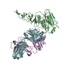
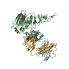
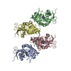
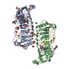
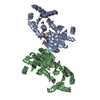
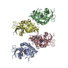
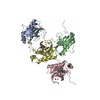
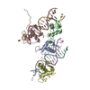
| Title | Crystal structure of the heterotrimeric PriSLX primase from S. solfataricus. | ||||||
 Components Components |
| ||||||
 Keywords Keywords | REPLICATION / Primase / DNA-dependent RNA polymerase / DNA replication | ||||||
| Function / homology |  Function and homology information Function and homology informationprimosome complex / DNA replication, synthesis of primer / DNA-directed RNA polymerase complex / Transferases; Transferring phosphorus-containing groups; Nucleotidyltransferases / DNA-directed RNA polymerase activity / 4 iron, 4 sulfur cluster binding / metal ion binding Similarity search - Function | ||||||
| Biological species |   Sulfolobus solfataricus (archaea) Sulfolobus solfataricus (archaea) | ||||||
| Method |  X-RAY DIFFRACTION / SYNCHROTRON / MOLECULAR REPLACEMENT / Resolution: 3.005 Å X-RAY DIFFRACTION / SYNCHROTRON / MOLECULAR REPLACEMENT / Resolution: 3.005 Å | ||||||
 Authors Authors | Pellegrini, L. / Holzer, S. | ||||||
| Funding support |  United Kingdom, 1items United Kingdom, 1items
| ||||||
 Citation Citation | Journal: Nat Commun / Year: 2017 Title: Primer synthesis by a eukaryotic-like archaeal primase is independent of its Fe-S cluster. Authors: Holzer, S. / Yan, J. / Kilkenny, M.L. / Bell, S.D. / Pellegrini, L. | ||||||
| History |
|
- Structure visualization
| Structure viewer | Molecule: MolmilJmol/JSmol |
|---|
- Downloads & links
-Download
| PDBx/mmCIF format | 5ofn.cif.gz | 145.4 KB | Display | PDBx/mmCIF format |
|---|---|---|---|---|
| PDB format | pdb5ofn.ent.gz | 108.8 KB | Display | PDB format |
| PDBx/mmJSON format | 5ofn.json.gz | Tree view | PDBx/mmJSON format | |
| Others |  Other downloads Other downloads |
-Validation report
| Arichive directory | https://data.pdbj.org/pub/pdb/validation_reports/of/5ofnftp://data.pdbj.org/pub/pdb/validation_reports/of/5ofn | HTTPS FTP |
|---|
-Related structure data
| Related structure data | 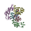 5of3C 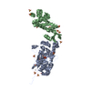 1zt2S S: Starting model for refinement C: citing same article ( |
|---|---|
| Similar structure data |


-Links
 PDBj
PDBj- Assembly
| Deposited unit | 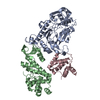
| ||||||||
|---|---|---|---|---|---|---|---|---|---|
| 1 |
| ||||||||
| Unit cell |
|
-Components
| #1: Protein | Mass: 37643.895 Da / Num. of mol.: 1 Source method: isolated from a genetically manipulated source Source: (gene. exp.) Sulfolobus solfataricus (strain ATCC 35092 / DSM 1617 / JCM 11322 / P2) (archaea)Strain: ATCC 35092 / DSM 1617 / JCM 11322 / P2 / Gene: priS, priA, SSO1048 / Production host:  References: UniProt: Q97Z83, Transferases; Transferring phosphorus-containing groups; Nucleotidyltransferases | ||||
|---|---|---|---|---|---|
| #2: Protein | Mass: 38173.637 Da / Num. of mol.: 2 Source method: isolated from a genetically manipulated source Details: Chain B is an engineered polypeptide. It contains the PriL residues of a chimeric construct spanning amino acids 1 to 211 of PriL fused to amino acids 42 to 154 of PriX. Source: (gene. exp.) Sulfolobus solfataricus (strain ATCC 35092 / DSM 1617 / JCM 11322 / P2) (archaea), (gene. exp.) Sulfolobus solfataricus (archaea)Strain: ATCC 35092 / DSM 1617 / JCM 11322 / P2 / Gene: priL, priB, SSO0557, C21_042 / Production host: #3: Chemical | ChemComp-ZN / |   Mass: 65.409 Da / Num. of mol.: 1 / Source method: isolated from a natural source / Formula: Zn Mass: 65.409 Da / Num. of mol.: 1 / Source method: isolated from a natural source / Formula: Zn#4: Water | ChemComp-HOH / |  Mass: 18.015 Da / Num. of mol.: 7 / Source method: isolated from a natural source / Formula: H2O Mass: 18.015 Da / Num. of mol.: 7 / Source method: isolated from a natural source / Formula: H2O |
-Experimental details
-Experiment
| Experiment | Method: X-RAY DIFFRACTION / Number of used crystals: 1 |
|---|
- Sample preparation
| Crystal | Density Matthews: 2.83 Å3/Da / Density % sol: 56.57 % |
|---|---|
| Crystal grow | Temperature: 291 K / Method: vapor diffusion, hanging drop Details: 12% PEG 3350, 200 mM CaCl2, 0.5% n-octyl-beta-D-glucoside |
-Data collection
| Diffraction | Mean temperature: 100 K |
|---|---|
| Diffraction source | Source: SYNCHROTRON / Site: Diamond / Beamline: I02 / Wavelength: 0.97949 Å |
| Detector | Type: DECTRIS PILATUS 6M-F / Detector: PIXEL / Date: Dec 12, 2015 |
| Radiation | Protocol: SINGLE WAVELENGTH / Monochromatic (M) / Laue (L): M / Scattering type: x-ray |
| Radiation wavelength | Wavelength: 0.97949 Å / Relative weight: 1 |
| Reflection | Resolution: 3.005→46.17 Å / Num. obs: 16582 / % possible obs: 98.63 % / Redundancy: 3.2 % / Biso Wilson estimate: 77 Å2 / CC1/2: 0.99 / Rmerge(I) obs: 0.1168 / Rpim(I) all: 0.07654 / Rsym value: 0.1402 / Net I/σ(I): 9.18 |
| Reflection shell | Resolution: 3.005→3.113 Å / Redundancy: 3.3 % / Rmerge(I) obs: 0.8095 / Mean I/σ(I) obs: 1.86 / Num. unique obs: 1556 / CC1/2: 0.535 / Rpim(I) all: 0.5204 / Rsym value: 0.9655 / % possible all: 95.17 |
- Processing
| Software |
| ||||||||||||||||||||||||||||||||||||||||||||||||||||||||||||||||||||||||||||||||||||
|---|---|---|---|---|---|---|---|---|---|---|---|---|---|---|---|---|---|---|---|---|---|---|---|---|---|---|---|---|---|---|---|---|---|---|---|---|---|---|---|---|---|---|---|---|---|---|---|---|---|---|---|---|---|---|---|---|---|---|---|---|---|---|---|---|---|---|---|---|---|---|---|---|---|---|---|---|---|---|---|---|---|---|---|---|---|
| Refinement | Method to determine structure: MOLECULAR REPLACEMENT Starting model: 1ZT2 Resolution: 3.005→46.17 Å / SU ML: 0.53 / Cross valid method: THROUGHOUT / σ(F): 0.04 / Phase error: 33.07
| ||||||||||||||||||||||||||||||||||||||||||||||||||||||||||||||||||||||||||||||||||||
| Solvent computation | Shrinkage radii: 0.9 Å / VDW probe radii: 1.11 Å | ||||||||||||||||||||||||||||||||||||||||||||||||||||||||||||||||||||||||||||||||||||
| Refinement step | Cycle: LAST / Resolution: 3.005→46.17 Å
| ||||||||||||||||||||||||||||||||||||||||||||||||||||||||||||||||||||||||||||||||||||
| Refine LS restraints |
| ||||||||||||||||||||||||||||||||||||||||||||||||||||||||||||||||||||||||||||||||||||
| LS refinement shell |
|
