| Entry | Database: PDB / ID: 5o54
|
|---|
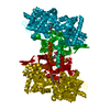
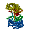
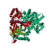
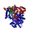
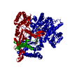
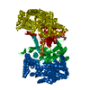
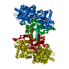
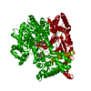
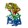
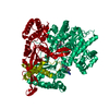
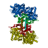
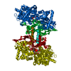
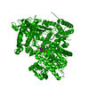
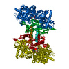
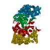
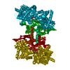
| Title | Glycogen Phosphorylase b in complex with 29a |
|---|
 Components Components | Glycogen phosphorylase, muscle form |
|---|
 Keywords Keywords | TRANSFERASE |
|---|
| Function / homology |  Function and homology information Function and homology information
glycogen phosphorylase / glycogen phosphorylase activity / glycogen catabolic process / skeletal muscle myofibril / pyridoxal phosphate binding / nucleotide bindingSimilarity search - Function Glycogen/starch/alpha-glucan phosphorylase / Phosphorylase pyridoxal-phosphate attachment site / Phosphorylase pyridoxal-phosphate attachment site. / Glycosyl transferase, family 35 / Carbohydrate phosphorylase / Glycogen Phosphorylase B; / Rossmann fold / 3-Layer(aba) Sandwich / Alpha BetaSimilarity search - Domain/homology |
|---|
| Biological species |   Oryctolagus cuniculus (rabbit) Oryctolagus cuniculus (rabbit) |
|---|
| Method |  X-RAY DIFFRACTION / FOURIER SYNTHESIS / Resolution: 2.45 Å X-RAY DIFFRACTION / FOURIER SYNTHESIS / Resolution: 2.45 Å |
|---|
 Authors Authors | Kyriakis, E. / Solovou, T.G.A. / Stravodimos, G.A. / Kantsadi, A.L. / Chatzileontiadou, D.S. / Skamnaki, V.T. / Leonidas, D.D. |
|---|
 Citation Citation | Journal: J. Med. Chem. / Year: 2017
Title: Nanomolar Inhibitors of Glycogen Phosphorylase Based on beta-d-Glucosaminyl Heterocycles: A Combined Synthetic, Enzyme Kinetic, and Protein Crystallography Study.
Authors: Bokor, E. / Kyriakis, E. / Solovou, T.G.A. / Koppany, C. / Kantsadi, A.L. / Szabo, K.E. / Szakacs, A. / Stravodimos, G.A. / Docsa, T. / Skamnaki, V.T. / Zographos, S.E. / Gergely, P. / ...Authors: Bokor, E. / Kyriakis, E. / Solovou, T.G.A. / Koppany, C. / Kantsadi, A.L. / Szabo, K.E. / Szakacs, A. / Stravodimos, G.A. / Docsa, T. / Skamnaki, V.T. / Zographos, S.E. / Gergely, P. / Leonidas, D.D. / Somsak, L. |
|---|
| History | | Deposition | May 31, 2017 | Deposition site: PDBE / Processing site: PDBE |
|---|
| Revision 1.0 | Sep 27, 2017 | Provider: repository / Type: Initial release |
|---|
| Revision 1.1 | Nov 29, 2017 | Group: Database references / Category: citation / citation_author
Item: _citation.journal_volume / _citation.page_first ..._citation.journal_volume / _citation.page_first / _citation.page_last / _citation.title / _citation_author.name |
|---|
| Revision 1.2 | Apr 9, 2025 | Group: Data collection / Database references / Structure summary
Category: chem_comp_atom / chem_comp_bond ...chem_comp_atom / chem_comp_bond / database_2 / pdbx_entry_details
Item: _database_2.pdbx_DOI / _database_2.pdbx_database_accession |
|---|
|
|---|
 Movie
Movie Controller


 Open data
 Basic information
 Structure visualization
 Downloads & links
 Other downloads
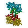


 PDBj
PDBj
 Assembly

 Mass: 247.142 Da / Num. of mol.: 1 / Source method: obtained synthetically / Formula: C8H10NO6P
 Mass: 306.317 Da / Num. of mol.: 1 / Source method: obtained synthetically / Formula: C14H18N4O4
 Mass: 348.206 Da / Num. of mol.: 1 / Source method: obtained synthetically / Formula: C10H13N4O8P
 Mass: 18.015 Da / Num. of mol.: 238 / Source method: isolated from a natural source / Formula: H2O
 Sample preparation
 Processing