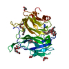
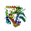
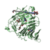
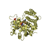
Entry Database : PDB / ID : 5o51Title AfRom2 CNH domain Rho guanyl nucleotide exchange factor (Rom2), putative Keywords / / / Function / homology Function Domain/homology Component
/ / / / / / / / / / / / / / / / / / / / / / / / / / / / / / / / Biological species Aspergillus fumigatus Af293 (mold)Method / / Resolution : 2.01 Å Authors Wei, W. / van Aalten, D. Funding support Organization Grant number Country Medical Research Council (United Kingdom)
Journal : To Be Published Title : Rom2 CNH domain from Aspergillus fumigatus is an atypical seven-bladed WD-40 proteinAuthors : Wei, W. / van Aalten, D. History Deposition May 31, 2017 Deposition site / Processing site Revision 1.0 Jun 13, 2018 Provider / Type Revision 1.1 Oct 16, 2019 Group / Category Revision 1.2 May 8, 2024 Group / Database references / Category / chem_comp_bond / database_2Item / _database_2.pdbx_database_accession
Show all Show less
 Movie
Movie Controller


 Open data
 Basic information
 Components
 Keywords
 Function and homology information
 X-RAY DIFFRACTION /
  Authors
 United Kingdom, 1items
  Citation
 Structure visualization
 Downloads & links
 Other downloads
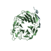


 PDBj
PDBj

 Assembly

 Mass: 18.015 Da / Num. of mol.: 72 / Source method: isolated from a natural source / Formula: H2O
 Sample preparation
 Processing