 Movie
Movie Controller

[English] 日本語
 Yorodumi
- PDB-5nx6: Crystal structure of 1,8-cineole synthase from Streptomyces clavu... -
+ Open data

- Basic information
| Entry | Database: PDB / ID: 5nx6 | ||||||
|---|---|---|---|---|---|---|---|
| Title | Crystal structure of 1,8-cineole synthase from Streptomyces clavuligerus in complex with 2-fluoroneryl diphosphate | ||||||
 Components Components | Pentalenene synthase | ||||||
 Keywords Keywords | LYASE / terpene synthase / 1 / 8-cineole / monoterpenoid / Eucalyptol | ||||||
| Function / homology |  Function and homology information Function and homology information1,8-cineole synthase / 1,8-cineole synthase activity / geranyl diphosphate metabolic process / terpene synthase activity / magnesium ion binding Similarity search - Function | ||||||
| Biological species |  Streptomyces clavuligerus (bacteria) Streptomyces clavuligerus (bacteria) | ||||||
| Method |  X-RAY DIFFRACTION / SYNCHROTRON / MOLECULAR REPLACEMENT / Resolution: 1.63 Å X-RAY DIFFRACTION / SYNCHROTRON / MOLECULAR REPLACEMENT / Resolution: 1.63 Å | ||||||
 Authors Authors | Karuppiah, V. / Leys, D. / Scrutton, N.S. | ||||||
| Funding support |  United Kingdom, 1items United Kingdom, 1items
| ||||||
 Citation Citation | Journal: ACS Catal / Year: 2017 Title: Structural Basis of Catalysis in the Bacterial Monoterpene Synthases Linalool Synthase and 1,8-Cineole Synthase. Authors: Karuppiah, V. / Ranaghan, K.E. / Leferink, N.G.H. / Johannissen, L.O. / Shanmugam, M. / Ni Cheallaigh, A. / Bennett, N.J. / Kearsey, L.J. / Takano, E. / Gardiner, J.M. / van der Kamp, M.W. / ...Authors: Karuppiah, V. / Ranaghan, K.E. / Leferink, N.G.H. / Johannissen, L.O. / Shanmugam, M. / Ni Cheallaigh, A. / Bennett, N.J. / Kearsey, L.J. / Takano, E. / Gardiner, J.M. / van der Kamp, M.W. / Hay, S. / Mulholland, A.J. / Leys, D. / Scrutton, N.S. | ||||||
| History |
|
- Structure visualization
| Structure viewer | Molecule: MolmilJmol/JSmol |
|---|
- Downloads & links
-Download
| PDBx/mmCIF format | 5nx6.cif.gz | 284.1 KB | Display | PDBx/mmCIF format |
|---|---|---|---|---|
| PDB format | pdb5nx6.ent.gz | 228.2 KB | Display | PDB format |
| PDBx/mmJSON format | 5nx6.json.gz | Tree view | PDBx/mmJSON format | |
| Others |  Other downloads Other downloads |
-Validation report
| Arichive directory | https://data.pdbj.org/pub/pdb/validation_reports/nx/5nx6ftp://data.pdbj.org/pub/pdb/validation_reports/nx/5nx6 | HTTPS FTP |
|---|
-Related structure data
| Related structure data |  5nx4C  5nx5C  5nx7C  1ps1S S: Starting model for refinement C: citing same article ( |
|---|---|
| Similar structure data |


-Links
 PDBj
PDBj
- Assembly
| Deposited unit | 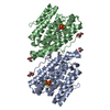
| ||||||||
|---|---|---|---|---|---|---|---|---|---|
| 1 |
| ||||||||
| Unit cell |
|
-Components
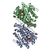
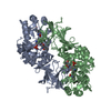
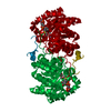
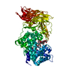
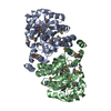
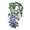
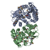
-Protein , 1 types, 2 molecules AB
| #1: Protein | Mass: 37774.359 Da / Num. of mol.: 2 Source method: isolated from a genetically manipulated source Source: (gene. exp.) Streptomyces clavuligerus (bacteria) / Gene: SCLAV_p0982, SSCG_00536 / Production host: |
|---|
-Non-polymers , 6 types, 821 molecules 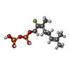

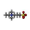


| #2: Chemical |  Mass: 332.200 Da / Num. of mol.: 2 Mass: 332.200 Da / Num. of mol.: 2Source method: isolated from a genetically manipulated source Formula: C10H19FO7P2 #3: Chemical | ChemComp-MG /  Mass: 24.305 Da / Num. of mol.: 4 / Source method: obtained synthetically / Formula: Mg Mass: 24.305 Da / Num. of mol.: 4 / Source method: obtained synthetically / Formula: Mg#4: Chemical |  Mass: 134.174 Da / Num. of mol.: 2 / Source method: obtained synthetically / Formula: C6H14O3 Mass: 134.174 Da / Num. of mol.: 2 / Source method: obtained synthetically / Formula: C6H14O3#5: Chemical |  Mass: 195.280 Da / Num. of mol.: 2 / Source method: obtained synthetically / Formula: C7H17NO3S Mass: 195.280 Da / Num. of mol.: 2 / Source method: obtained synthetically / Formula: C7H17NO3S#6: Chemical |  Mass: 92.094 Da / Num. of mol.: 2 / Source method: obtained synthetically / Formula: C3H8O3 Mass: 92.094 Da / Num. of mol.: 2 / Source method: obtained synthetically / Formula: C3H8O3#7: Water | ChemComp-HOH / | Mass: 18.015 Da / Num. of mol.: 809 / Source method: isolated from a natural source / Formula: H2O |
|---|
-Experimental details
-Experiment
| Experiment | Method: X-RAY DIFFRACTION / Number of used crystals: 1 |
|---|
- Sample preparation
| Crystal | Density Matthews: 3.2 Å3/Da / Density % sol: 61.57 % |
|---|---|
| Crystal grow | Temperature: 277.15 K / Method: vapor diffusion, sitting drop Details: 90mM of LiNaK (0.3M lithium sulphate, 0.3M sodium sulphate, 0.3 M potassium sulphate), 0.1M of buffer system 4 (1M MOPSO, 1M Bis-Tris) pH 6.5 and 50% precipitant mix 8 (10% PEG 20000, 50% ...Details: 90mM of LiNaK (0.3M lithium sulphate, 0.3M sodium sulphate, 0.3 M potassium sulphate), 0.1M of buffer system 4 (1M MOPSO, 1M Bis-Tris) pH 6.5 and 50% precipitant mix 8 (10% PEG 20000, 50% trimethyl propane, 2% NDSB 195) |
-Data collection
| Diffraction | Mean temperature: 100 K |
|---|---|
| Diffraction source | Source: SYNCHROTRON / Site: Diamond / Beamline: I04 / Wavelength: 0.99 Å |
| Detector | Type: DECTRIS PILATUS 6M-F / Detector: PIXEL / Date: May 5, 2016 |
| Radiation | Protocol: SINGLE WAVELENGTH / Monochromatic (M) / Laue (L): M / Scattering type: x-ray |
| Radiation wavelength | Wavelength: 0.99 Å / Relative weight: 1 |
| Reflection | Resolution: 1.63→32.27 Å / Num. obs: 107492 / % possible obs: 96 % / Redundancy: 1.8 % / CC1/2: 0.987 / Rmerge(I) obs: 0.09 / Rpim(I) all: 0.09 / Net I/σ(I): 6.7 |
| Reflection shell | Resolution: 1.63→1.66 Å / Redundancy: 1.8 % / Rmerge(I) obs: 0.639 / Mean I/σ(I) obs: 1.2 / Num. unique obs: 5258 / CC1/2: 0.479 / Rpim(I) all: 0.639 / % possible all: 94.6 |
- Processing
| Software |
| |||||||||||||||||||||||||||||||||||||||||||||||||||||||||||||||||||||||||||||||||||||||||||||||||||||||||||||||||||||||||||||||||||||||||||||||||||||||||||||||||||||||||||||||||||||||||||||||||||||||||||||||||||||||||
|---|---|---|---|---|---|---|---|---|---|---|---|---|---|---|---|---|---|---|---|---|---|---|---|---|---|---|---|---|---|---|---|---|---|---|---|---|---|---|---|---|---|---|---|---|---|---|---|---|---|---|---|---|---|---|---|---|---|---|---|---|---|---|---|---|---|---|---|---|---|---|---|---|---|---|---|---|---|---|---|---|---|---|---|---|---|---|---|---|---|---|---|---|---|---|---|---|---|---|---|---|---|---|---|---|---|---|---|---|---|---|---|---|---|---|---|---|---|---|---|---|---|---|---|---|---|---|---|---|---|---|---|---|---|---|---|---|---|---|---|---|---|---|---|---|---|---|---|---|---|---|---|---|---|---|---|---|---|---|---|---|---|---|---|---|---|---|---|---|---|---|---|---|---|---|---|---|---|---|---|---|---|---|---|---|---|---|---|---|---|---|---|---|---|---|---|---|---|---|---|---|---|---|---|---|---|---|---|---|---|---|---|---|---|---|---|---|---|---|
| Refinement | Method to determine structure: MOLECULAR REPLACEMENT Starting model: 1PS1 Resolution: 1.63→32.264 Å / SU ML: 0.19 / Cross valid method: FREE R-VALUE / σ(F): 1.96 / Phase error: 20.37
| |||||||||||||||||||||||||||||||||||||||||||||||||||||||||||||||||||||||||||||||||||||||||||||||||||||||||||||||||||||||||||||||||||||||||||||||||||||||||||||||||||||||||||||||||||||||||||||||||||||||||||||||||||||||||
| Solvent computation | Shrinkage radii: 0.9 Å / VDW probe radii: 1.11 Å | |||||||||||||||||||||||||||||||||||||||||||||||||||||||||||||||||||||||||||||||||||||||||||||||||||||||||||||||||||||||||||||||||||||||||||||||||||||||||||||||||||||||||||||||||||||||||||||||||||||||||||||||||||||||||
| Refinement step | Cycle: LAST / Resolution: 1.63→32.264 Å
| |||||||||||||||||||||||||||||||||||||||||||||||||||||||||||||||||||||||||||||||||||||||||||||||||||||||||||||||||||||||||||||||||||||||||||||||||||||||||||||||||||||||||||||||||||||||||||||||||||||||||||||||||||||||||
| Refine LS restraints |
| |||||||||||||||||||||||||||||||||||||||||||||||||||||||||||||||||||||||||||||||||||||||||||||||||||||||||||||||||||||||||||||||||||||||||||||||||||||||||||||||||||||||||||||||||||||||||||||||||||||||||||||||||||||||||
| LS refinement shell |
| |||||||||||||||||||||||||||||||||||||||||||||||||||||||||||||||||||||||||||||||||||||||||||||||||||||||||||||||||||||||||||||||||||||||||||||||||||||||||||||||||||||||||||||||||||||||||||||||||||||||||||||||||||||||||
| Refinement TLS params. | Method: refined / Refine-ID: X-RAY DIFFRACTION
| |||||||||||||||||||||||||||||||||||||||||||||||||||||||||||||||||||||||||||||||||||||||||||||||||||||||||||||||||||||||||||||||||||||||||||||||||||||||||||||||||||||||||||||||||||||||||||||||||||||||||||||||||||||||||
| Refinement TLS group |
|
