 Movie
Movie Controller

[English] 日本語
 Yorodumi
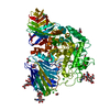
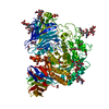
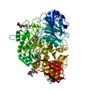
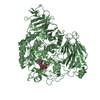
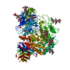
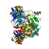
- PDB-5nn5: Crystal structure of human lysosomal acid-alpha-glucosidase, GAA,... -
+ Open data

- Basic information
| Entry | Database: PDB / ID: 5nn5 | |||||||||
|---|---|---|---|---|---|---|---|---|---|---|
| Title | Crystal structure of human lysosomal acid-alpha-glucosidase, GAA, in complex with 1-deoxynojirimycin | |||||||||
 Components Components | Lysosomal alpha-glucosidase | |||||||||
 Keywords Keywords | HYDROLASE / glycoside hydrolase / lysosome / glycogen catabolism / Pompe disease | |||||||||
| Function / homology |  Function and homology information Function and homology information: / autolysosome lumen / maltose metabolic process / glucan 1,6-alpha-glucosidase activity / alpha-glucosidase activity / sucrose metabolic process / Glycogen storage disease type II (GAA) / alpha-glucosidase / alpha-1,4-glucosidase activity / neuromuscular process controlling posture ...: / autolysosome lumen / maltose metabolic process / glucan 1,6-alpha-glucosidase activity / alpha-glucosidase activity / sucrose metabolic process / Glycogen storage disease type II (GAA) / alpha-glucosidase / alpha-1,4-glucosidase activity / neuromuscular process controlling posture / glycophagy / diaphragm contraction / tissue development / regulation of the force of heart contraction / glycogen catabolic process / neuromuscular process controlling balance / aorta development / azurophil granule membrane / muscle cell cellular homeostasis / lysosome organization / Glycogen breakdown (glycogenolysis) / tertiary granule membrane / ficolin-1-rich granule membrane / heart morphogenesis / cardiac muscle contraction / lysosomal lumen / locomotory behavior / glucose metabolic process / carbohydrate binding / lysosome / lysosomal membrane / Neutrophil degranulation / extracellular exosome / membrane / plasma membrane Similarity search - Function | |||||||||
| Biological species |  Homo sapiens (human) Homo sapiens (human) | |||||||||
| Method |  X-RAY DIFFRACTION / SYNCHROTRON / FOURIER SYNTHESIS / Resolution: 2 Å X-RAY DIFFRACTION / SYNCHROTRON / FOURIER SYNTHESIS / Resolution: 2 Å | |||||||||
 Authors Authors | Roig-Zamboni, V. / Cobucci-Ponzano, B. / Iacono, R. / Ferrara, M.C. / Germany, S. / Parenti, G. / Bourne, Y. / Moracci, M. | |||||||||
 Citation Citation | Journal: Nat Commun / Year: 2017 Title: Structure of human lysosomal acid alpha-glucosidase-a guide for the treatment of Pompe disease. Authors: Roig-Zamboni, V. / Cobucci-Ponzano, B. / Iacono, R. / Ferrara, M.C. / Germany, S. / Bourne, Y. / Parenti, G. / Moracci, M. / Sulzenbacher, G. | |||||||||
| History |
|
- Structure visualization
| Structure viewer | Molecule: MolmilJmol/JSmol |
|---|
- Downloads & links
-Download
| PDBx/mmCIF format | 5nn5.cif.gz | 359.9 KB | Display | PDBx/mmCIF format |
|---|---|---|---|---|
| PDB format | pdb5nn5.ent.gz | 287.1 KB | Display | PDB format |
| PDBx/mmJSON format | 5nn5.json.gz | Tree view | PDBx/mmJSON format | |
| Others |  Other downloads Other downloads |
-Validation report
| Arichive directory | https://data.pdbj.org/pub/pdb/validation_reports/nn/5nn5ftp://data.pdbj.org/pub/pdb/validation_reports/nn/5nn5 | HTTPS FTP |
|---|
-Related structure data
| Related structure data |  5nn3SC  5nn4C  5nn6C  5nn8C S: Starting model for refinement C: citing same article ( |
|---|---|
| Similar structure data |


-Links
 PDBj
PDBj


- Assembly
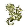
| Deposited unit | 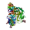
| ||||||||
|---|---|---|---|---|---|---|---|---|---|
| 1 |
| ||||||||
| Unit cell |
|
-Components
-Protein , 1 types, 1 molecules A
| #1: Protein | Mass: 96978.633 Da / Num. of mol.: 1 Source method: isolated from a genetically manipulated source Source: (gene. exp.) Homo sapiens (human) / Gene: GAA / Cell (production host): Ovary cells / Production host:   Cricetulus griseus (Chinese hamster) / References: UniProt: P10253, alpha-glucosidase Cricetulus griseus (Chinese hamster) / References: UniProt: P10253, alpha-glucosidase |
|---|
-Sugars , 5 types, 6 molecules 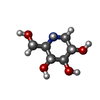
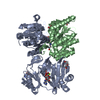
| #2: Polysaccharide | Source method: isolated from a genetically manipulated source #3: Polysaccharide | beta-D-mannopyranose-(1-4)-2-acetamido-2-deoxy-beta-D-glucopyranose-(1-4)-2-acetamido-2-deoxy-beta- ...beta-D-mannopyranose-(1-4)-2-acetamido-2-deoxy-beta-D-glucopyranose-(1-4)-2-acetamido-2-deoxy-beta-D-glucopyranose | Source method: isolated from a genetically manipulated source #4: Polysaccharide | 2-acetamido-2-deoxy-beta-D-glucopyranose-(1-4)-2-acetamido-2-deoxy-beta-D-glucopyranose | Source method: isolated from a genetically manipulated source #5: Polysaccharide | alpha-D-mannopyranose-(1-3)-beta-D-mannopyranose-(1-4)-2-acetamido-2-deoxy-beta-D-glucopyranose-(1- ...alpha-D-mannopyranose-(1-3)-beta-D-mannopyranose-(1-4)-2-acetamido-2-deoxy-beta-D-glucopyranose-(1-4)-2-acetamido-2-deoxy-beta-D-glucopyranose | Source method: isolated from a genetically manipulated source #6: Sugar | ChemComp-NOJ / |  Type: D-saccharide / Mass: 163.172 Da / Num. of mol.: 1 / Source method: obtained synthetically / Formula: C6H13NO4 Type: D-saccharide / Mass: 163.172 Da / Num. of mol.: 1 / Source method: obtained synthetically / Formula: C6H13NO4 |
|---|
-Non-polymers , 4 types, 790 molecules 


| #7: Chemical | ChemComp-SO4 /  Mass: 96.063 Da / Num. of mol.: 4 / Source method: obtained synthetically / Formula: SO4 Mass: 96.063 Da / Num. of mol.: 4 / Source method: obtained synthetically / Formula: SO4#8: Chemical | ChemComp-CL /  Mass: 35.453 Da / Num. of mol.: 6 / Source method: obtained synthetically / Formula: Cl Mass: 35.453 Da / Num. of mol.: 6 / Source method: obtained synthetically / Formula: Cl#9: Chemical | ChemComp-EDO /  Mass: 62.068 Da / Num. of mol.: 7 / Source method: obtained synthetically / Formula: C2H6O2 Mass: 62.068 Da / Num. of mol.: 7 / Source method: obtained synthetically / Formula: C2H6O2#10: Water | ChemComp-HOH / | Mass: 18.015 Da / Num. of mol.: 773 / Source method: isolated from a natural source / Formula: H2O |
|---|
-Details
| Has protein modification | Y |
|---|
-Experimental details
-Experiment
| Experiment | Method: X-RAY DIFFRACTION / Number of used crystals: 1 |
|---|
- Sample preparation
| Crystal | Density Matthews: 3.39 Å3/Da / Density % sol: 63.73 % |
|---|---|
| Crystal grow | Temperature: 293 K / Method: vapor diffusion, hanging drop / pH: 7 Details: 1.9 M ammonium sulphate, 0.1 M HEPES pH 7.0, 2% v/v PEG400 PH range: 7 |
-Data collection
| Diffraction | Mean temperature: 100 K |
|---|---|
| Diffraction source | Source: SYNCHROTRON / Site: ESRF  / Beamline: ID23-2 / Wavelength: 0.8726 Å / Beamline: ID23-2 / Wavelength: 0.8726 Å |
| Detector | Type: DECTRIS PILATUS3 6M / Detector: PIXEL / Date: Apr 18, 2014 |
| Radiation | Monochromator: 0.8726 / Protocol: SINGLE WAVELENGTH / Monochromatic (M) / Laue (L): M / Scattering type: x-ray |
| Radiation wavelength | Wavelength: 0.8726 Å / Relative weight: 1 |
| Reflection | Resolution: 2→53.86 Å / Num. obs: 83609 / % possible obs: 95.3 % / Redundancy: 3.1 % / Biso Wilson estimate: 14.22 Å2 / CC1/2: 0.965 / Rmerge(I) obs: 0.169 / Rpim(I) all: 0.125 / Net I/σ(I): 6.2 |
| Reflection shell | Resolution: 2→2.04 Å / Redundancy: 3 % / Rmerge(I) obs: 0.549 / Mean I/σ(I) obs: 2 / Num. unique obs: 4552 / CC1/2: 0.675 / Rpim(I) all: 0.426 / % possible all: 95.5 |
- Processing
| Software |
| ||||||||||||||||||||||||||||||||||||||||||||||||||||||||||||||||||||||||||||||||||||||||||||||||||||||||||||||||||||||||||||||||||||||||||||||||||||||||||||||||||||||||||||||||||||||
|---|---|---|---|---|---|---|---|---|---|---|---|---|---|---|---|---|---|---|---|---|---|---|---|---|---|---|---|---|---|---|---|---|---|---|---|---|---|---|---|---|---|---|---|---|---|---|---|---|---|---|---|---|---|---|---|---|---|---|---|---|---|---|---|---|---|---|---|---|---|---|---|---|---|---|---|---|---|---|---|---|---|---|---|---|---|---|---|---|---|---|---|---|---|---|---|---|---|---|---|---|---|---|---|---|---|---|---|---|---|---|---|---|---|---|---|---|---|---|---|---|---|---|---|---|---|---|---|---|---|---|---|---|---|---|---|---|---|---|---|---|---|---|---|---|---|---|---|---|---|---|---|---|---|---|---|---|---|---|---|---|---|---|---|---|---|---|---|---|---|---|---|---|---|---|---|---|---|---|---|---|---|---|---|
| Refinement | Method to determine structure: FOURIER SYNTHESIS Starting model: 5NN3 Resolution: 2→53.86 Å / Cor.coef. Fo:Fc: 0.928 / Cor.coef. Fo:Fc free: 0.907 / SU B: 6.245 / SU ML: 0.09 / Cross valid method: THROUGHOUT / ESU R: 0.152 / ESU R Free: 0.141 / Details: HYDROGENS HAVE BEEN ADDED IN THE RIDING POSITIONS
| ||||||||||||||||||||||||||||||||||||||||||||||||||||||||||||||||||||||||||||||||||||||||||||||||||||||||||||||||||||||||||||||||||||||||||||||||||||||||||||||||||||||||||||||||||||||
| Solvent computation | Ion probe radii: 0.7 Å / Shrinkage radii: 0.7 Å / VDW probe radii: 1 Å | ||||||||||||||||||||||||||||||||||||||||||||||||||||||||||||||||||||||||||||||||||||||||||||||||||||||||||||||||||||||||||||||||||||||||||||||||||||||||||||||||||||||||||||||||||||||
| Displacement parameters | Biso mean: 19.493 Å2
| ||||||||||||||||||||||||||||||||||||||||||||||||||||||||||||||||||||||||||||||||||||||||||||||||||||||||||||||||||||||||||||||||||||||||||||||||||||||||||||||||||||||||||||||||||||||
| Refinement step | Cycle: 1 / Resolution: 2→53.86 Å
| ||||||||||||||||||||||||||||||||||||||||||||||||||||||||||||||||||||||||||||||||||||||||||||||||||||||||||||||||||||||||||||||||||||||||||||||||||||||||||||||||||||||||||||||||||||||
| Refine LS restraints |
|