 Movie
Movie Controller

[English] 日本語
 Yorodumi
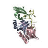
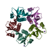
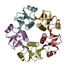
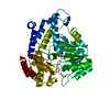
- PDB-5new: RNA-RNA base stacking in the crystal structure of an Hfq6:RNA dimer -
+ Open data

- Basic information
| Entry | Database: PDB / ID: 5new | ||||||
|---|---|---|---|---|---|---|---|
| Title | RNA-RNA base stacking in the crystal structure of an Hfq6:RNA dimer | ||||||
 Components Components |
| ||||||
 Keywords Keywords | RNA / Hfq sRNA RNA-RNA interaction base stacking | ||||||
| Function / homology |  Function and homology information Function and homology informationregulation of translation, ncRNA-mediated / regulation of RNA stability / regulation of DNA-templated transcription / RNA binding / cytosol Similarity search - Function | ||||||
| Biological species |  | ||||||
| Method |  X-RAY DIFFRACTION / SYNCHROTRON / Resolution: 2.511 Å X-RAY DIFFRACTION / SYNCHROTRON / Resolution: 2.511 Å | ||||||
 Authors Authors | Schulz, E.C. / Barabas, O. | ||||||
 Citation Citation | Journal: Sci Rep / Year: 2017 Title: Intermolecular base stacking mediates RNA-RNA interaction in a crystal structure of the RNA chaperone Hfq. Authors: Schulz, E.C. / Seiler, M. / Zuliani, C. / Voigt, F. / Rybin, V. / Pogenberg, V. / Mucke, N. / Wilmanns, M. / Gibson, T.J. / Barabas, O. | ||||||
| History |
|
- Structure visualization
| Structure viewer | Molecule: MolmilJmol/JSmol |
|---|
- Downloads & links
-Download
| PDBx/mmCIF format | 5new.cif.gz | 45.9 KB | Display | PDBx/mmCIF format |
|---|---|---|---|---|
| PDB format | pdb5new.ent.gz | 31.8 KB | Display | PDB format |
| PDBx/mmJSON format | 5new.json.gz | Tree view | PDBx/mmJSON format | |
| Others |  Other downloads Other downloads |
-Validation report
| Arichive directory | https://data.pdbj.org/pub/pdb/validation_reports/ne/5newftp://data.pdbj.org/pub/pdb/validation_reports/ne/5new | HTTPS FTP |
|---|
-Related structure data
| Similar structure data |
|---|


-Links
 PDBj
PDBj

- Assembly
| Deposited unit | 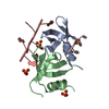
| ||||||||
|---|---|---|---|---|---|---|---|---|---|
| 1 | 
| ||||||||
| Unit cell |
|
-Components
| #1: Protein | Mass: 11179.354 Da / Num. of mol.: 2 Source method: isolated from a genetically manipulated source Source: (gene. exp.) #2: RNA chain | | Mass: 1930.277 Da / Num. of mol.: 1 / Source method: obtained synthetically / Source: (synth.) #3: RNA chain | | Mass: 567.374 Da / Num. of mol.: 1 / Source method: isolated from a natural source / Source: (natural) #4: Chemical | ChemComp-SO4 /   Mass: 96.063 Da / Num. of mol.: 5 / Source method: obtained synthetically / Formula: SO4 Mass: 96.063 Da / Num. of mol.: 5 / Source method: obtained synthetically / Formula: SO4#5: Water | ChemComp-HOH / |  Mass: 18.015 Da / Num. of mol.: 44 / Source method: isolated from a natural source / Formula: H2O Mass: 18.015 Da / Num. of mol.: 44 / Source method: isolated from a natural source / Formula: H2O |
|---|
-Experimental details
-Experiment
| Experiment | Method: X-RAY DIFFRACTION / Number of used crystals: 1 |
|---|
- Sample preparation
| Crystal | Density Matthews: 1.97 Å3/Da / Density % sol: 37.61 % |
|---|---|
| Crystal grow | Temperature: 293 K / Method: vapor diffusion, hanging drop / pH: 4.2 Details: 0.1 M phosphate-citrate buffer pH 4.2, 27% PEG 1000, and 0.2 M LiSO4 |
-Data collection
| Diffraction | Mean temperature: 100 K |
|---|---|
| Diffraction source | Source: SYNCHROTRON / Site: ESRF  / Beamline: BM30A / Wavelength: 0.979681 Å / Beamline: BM30A / Wavelength: 0.979681 Å |
| Detector | Type: ADSC QUANTUM 315 / Detector: CCD / Date: Dec 4, 2011 |
| Radiation | Protocol: SINGLE WAVELENGTH / Monochromatic (M) / Laue (L): M / Scattering type: x-ray |
| Radiation wavelength | Wavelength: 0.979681 Å / Relative weight: 1 |
| Reflection | Resolution: 2.51→40.6 Å / Num. obs: 6816 / % possible obs: 96.8 % / Redundancy: 4.6 % / Net I/σ(I): 15.03 |
- Processing
| Software |
| ||||||||||||||||||||||||
|---|---|---|---|---|---|---|---|---|---|---|---|---|---|---|---|---|---|---|---|---|---|---|---|---|---|
| Refinement | Resolution: 2.511→40.6 Å / SU ML: 0.29 / Cross valid method: FREE R-VALUE / σ(F): 1.99 / Phase error: 28.65
| ||||||||||||||||||||||||
| Solvent computation | Shrinkage radii: 0.9 Å / VDW probe radii: 1.11 Å / Bsol: 44.329 Å2 / ksol: 0.392 e/Å3 | ||||||||||||||||||||||||
| Displacement parameters |
| ||||||||||||||||||||||||
| Refinement step | Cycle: LAST / Resolution: 2.511→40.6 Å
| ||||||||||||||||||||||||
| Refine LS restraints |
| ||||||||||||||||||||||||
| LS refinement shell |
|
