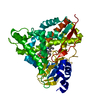
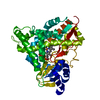
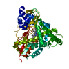
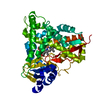
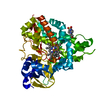
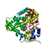
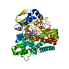
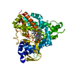
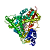
[English] 日本語
Yorodumi
- PDB-5ncb: Crystal structure of Amycolatopsis cytochrome P450 GcoA in comple... -

+
Open data


ID or keywords:

Loading...

-
Basic information

Entry
Database: PDB / ID: 5ncb
TitleCrystal structure of Amycolatopsis cytochrome P450 GcoA in complex with guaiacol.
ComponentsCytochrome P450
KeywordsOXIDOREDUCTASE / Cytochrome / P450 / guaiacol / lignin / CYP255A / Amycolatopsis / heme / haem
Function / homology
Function and homology information


cholest-4-en-3-one 26-monooxygenase activity / steroid hydroxylase activity / cholesterol catabolic process / iron ion binding / heme binding
Similarity search - Function
Cytochrome P450, B-class / Cytochrome P450, conserved site / Cytochrome P450 cysteine heme-iron ligand signature. / Cytochrome P450 / Cytochrome P450 superfamily / Cytochrome P450
Similarity search - Domain/homology
PROTOPORPHYRIN IX CONTAINING FE / Guaiacol / Cytochrome P450
Similarity search - Component
Biological speciesAmycolatopsis sp. ATCC 39116 (bacteria)
MethodX-RAY DIFFRACTION / SYNCHROTRON / SAD / Resolution: 1.44 Å
AuthorsMallinson, S.J.B. / Johnson, C.W. / Neidle, E.L. / Beckham, G.T. / McGeehan, J.E.
Funding support United Kingdom, United States, 3items
OrganizationGrant numberCountry
Biotechnology and Biological Sciences Research CouncilBB/P0119818/1 United Kingdom
Biotechnology and Biological Sciences Research CouncilBB/L001926/1 United Kingdom
Department of Energy (DOE, United States)DE-AC36-08GO28308 United States
CitationJournal: Nat Commun / Year: 2018
Title: A promiscuous cytochrome P450 aromatic O-demethylase for lignin bioconversion.
Authors: Mallinson, S.J.B. / Machovina, M.M. / Silveira, R.L. / Garcia-Borras, M. / Gallup, N. / Johnson, C.W. / Allen, M.D. / Skaf, M.S. / Crowley, M.F. / Neidle, E.L. / Houk, K.N. / Beckham, G.T. / ...Authors: Mallinson, S.J.B. / Machovina, M.M. / Silveira, R.L. / Garcia-Borras, M. / Gallup, N. / Johnson, C.W. / Allen, M.D. / Skaf, M.S. / Crowley, M.F. / Neidle, E.L. / Houk, K.N. / Beckham, G.T. / DuBois, J.L. / McGeehan, J.E.
History
DepositionMar 3, 2017Deposition site: PDBE / Processing site: PDBE
Revision 1.0Jul 4, 2018Provider: repository / Type: Initial release
Revision 1.1Jul 11, 2018Group: Data collection / Database references / Category: citation
Item: _citation.journal_volume / _citation.page_first ..._citation.journal_volume / _citation.page_first / _citation.page_last / _citation.pdbx_database_id_PubMed / _citation.title
Revision 1.2Mar 30, 2022Group: Author supporting evidence / Database references / Derived calculations
Category: database_2 / pdbx_audit_support / struct_conn
Item: _database_2.pdbx_DOI / _database_2.pdbx_database_accession ..._database_2.pdbx_DOI / _database_2.pdbx_database_accession / _pdbx_audit_support.funding_organization / _struct_conn.conn_type_id / _struct_conn.id / _struct_conn.pdbx_dist_value / _struct_conn.pdbx_leaving_atom_flag / _struct_conn.ptnr1_auth_comp_id / _struct_conn.ptnr1_auth_seq_id / _struct_conn.ptnr1_label_atom_id / _struct_conn.ptnr1_label_comp_id / _struct_conn.ptnr1_label_seq_id / _struct_conn.ptnr2_auth_comp_id / _struct_conn.ptnr2_auth_seq_id / _struct_conn.ptnr2_label_asym_id / _struct_conn.ptnr2_label_atom_id / _struct_conn.ptnr2_label_comp_id / _struct_conn.ptnr2_label_seq_id

-
Structure visualization

Structure viewerMolecule:
MolmilJmol/JSmol

Downloads & links

-
Assembly

Deposited unit
A: Cytochrome P450
hetero molecules


Theoretical massNumber of molelcules
Total (without water)46,5693
Polymers45,8281
Non-polymers7412
Water5,459303
1


  • Idetical with deposited unit
  • defined by author&software
  • Evidence: SAXS
TypeNameSymmetry operationNumber
identity operation1_555x,y,z1
Buried area1230 Å2
ΔGint-25 kcal/mol
Surface area16270 Å2
MethodPISA
Unit cell
Length a, b, c (Å)104.080, 104.080, 115.754
Angle α, β, γ (deg.)90.00, 90.00, 90.00
Int Tables number96
Space group name H-MP43212
Components on special symmetry positions
IDModelComponents
11A-892-

HOH

-
Components

#1: Protein Cytochrome P450


Mass: 45827.934 Da / Num. of mol.: 1
Source method: isolated from a genetically manipulated source
Source: (gene. exp.) Amycolatopsis sp. ATCC 39116 (bacteria)
Gene: WP_020419855 / Production host: Escherichia coli (E. coli) / References: UniProt: A0A076MY51
#2: Chemical ChemComp-HEM / PROTOPORPHYRIN IX CONTAINING FE / HEME


Mass: 616.487 Da / Num. of mol.: 1 / Source method: obtained synthetically / Formula: C34H32FeN4O4
#3: Chemical ChemComp-JZ3 / Guaiacol / 2-methoxyphenol


Mass: 124.137 Da / Num. of mol.: 1 / Source method: obtained synthetically / Formula: C7H8O2
#4: Water ChemComp-HOH / water


Mass: 18.015 Da / Num. of mol.: 303 / Source method: isolated from a natural source / Formula: H2O

-
Experimental details

-
Experiment

ExperimentMethod: X-RAY DIFFRACTION / Number of used crystals: 1

-
Sample preparation

CrystalDensity Matthews: 3.42 Å3/Da / Density % sol: 64.04 %
Crystal growTemperature: 279 K / Method: vapor diffusion, hanging drop / Details: Sodium malonate, HEPES, guaiacol.

-
Data collection

DiffractionMean temperature: 100 K
Diffraction sourceSource: SYNCHROTRON / Site: Diamond / Beamline: I04 / Wavelength: 0.9795 Å
DetectorType: DECTRIS PILATUS 6M-F / Detector: PIXEL / Date: Jul 12, 2016
RadiationProtocol: SINGLE WAVELENGTH / Monochromatic (M) / Laue (L): M / Scattering type: x-ray
Radiation wavelengthWavelength: 0.9795 Å / Relative weight: 1
ReflectionResolution: 1.44→115.75 Å / Num. obs: 114601 / % possible obs: 99.7 % / Redundancy: 10.2 % / CC1/2: 0.998 / Rmerge(I) obs: 0.073 / Rpim(I) all: 0.024 / Net I/σ(I): 21
Reflection shellResolution: 1.44→1.46 Å / Redundancy: 8.6 % / Rmerge(I) obs: 0.931 / Mean I/σ(I) obs: 2.1 / Num. unique obs: 5611 / CC1/2: 0.794 / Rpim(I) all: 0.333 / % possible all: 99.6

-
Processing

Software
NameVersionClassification
REFMAC5.8.0158refinement
xia2data reduction
xia2data scaling
CRANK2phasing
RefinementMethod to determine structure: SAD / Resolution: 1.44→77.39 Å / Cor.coef. Fo:Fc: 0.981 / Cor.coef. Fo:Fc free: 0.978 / SU B: 1.773 / SU ML: 0.029 / Cross valid method: THROUGHOUT / ESU R: 0.04 / ESU R Free: 0.04 / Details: HYDROGENS HAVE BEEN ADDED IN THE RIDING POSITIONS
RfactorNum. reflection% reflectionSelection details
Rfree0.15141 5812 5.1 %RANDOM
Rwork0.12773 ---
obs0.12892 108389 99.46 %-
Solvent computationIon probe radii: 0.8 Å / Shrinkage radii: 0.8 Å / VDW probe radii: 1.4 Å
Displacement parametersBiso mean: 23.622 Å2
Baniso -1Baniso -2Baniso -3
1--0.41 Å20 Å20 Å2
2---0.41 Å20 Å2
3---0.81 Å2
Refinement stepCycle: 1 / Resolution: 1.44→77.39 Å
ProteinNucleic acidLigandSolventTotal
Num. atoms3164 0 52 303 3519
Refine LS restraints
Refine-IDTypeDev idealDev ideal targetNumber
X-RAY DIFFRACTIONr_bond_refined_d0.0150.0193357
X-RAY DIFFRACTIONr_bond_other_d0.0020.022991
X-RAY DIFFRACTIONr_angle_refined_deg1.711.9774606
X-RAY DIFFRACTIONr_angle_other_deg1.07636918
X-RAY DIFFRACTIONr_dihedral_angle_1_deg5.7615410
X-RAY DIFFRACTIONr_dihedral_angle_2_deg32.37723.558163
X-RAY DIFFRACTIONr_dihedral_angle_3_deg11.1815472
X-RAY DIFFRACTIONr_dihedral_angle_4_deg21.3991527
X-RAY DIFFRACTIONr_chiral_restr0.160.2487
X-RAY DIFFRACTIONr_gen_planes_refined0.0130.0213824
X-RAY DIFFRACTIONr_gen_planes_other0.0040.02719
X-RAY DIFFRACTIONr_nbd_refined
X-RAY DIFFRACTIONr_nbd_other
X-RAY DIFFRACTIONr_nbtor_refined
X-RAY DIFFRACTIONr_nbtor_other
X-RAY DIFFRACTIONr_xyhbond_nbd_refined
X-RAY DIFFRACTIONr_xyhbond_nbd_other
X-RAY DIFFRACTIONr_metal_ion_refined
X-RAY DIFFRACTIONr_metal_ion_other
X-RAY DIFFRACTIONr_symmetry_vdw_refined
X-RAY DIFFRACTIONr_symmetry_vdw_other
X-RAY DIFFRACTIONr_symmetry_hbond_refined
X-RAY DIFFRACTIONr_symmetry_hbond_other
X-RAY DIFFRACTIONr_symmetry_metal_ion_refined
X-RAY DIFFRACTIONr_symmetry_metal_ion_other
X-RAY DIFFRACTIONr_mcbond_it1.5792.1471631
X-RAY DIFFRACTIONr_mcbond_other1.5682.1441630
X-RAY DIFFRACTIONr_mcangle_it2.0083.2292041
X-RAY DIFFRACTIONr_mcangle_other2.0093.2312042
X-RAY DIFFRACTIONr_scbond_it2.7022.4781726
X-RAY DIFFRACTIONr_scbond_other2.7012.4781727
X-RAY DIFFRACTIONr_scangle_it
X-RAY DIFFRACTIONr_scangle_other3.2023.6132565
X-RAY DIFFRACTIONr_long_range_B_refined3.2426.4883888
X-RAY DIFFRACTIONr_long_range_B_other3.07226.143826
X-RAY DIFFRACTIONr_rigid_bond_restr2.74336347
X-RAY DIFFRACTIONr_sphericity_free20.8275202
X-RAY DIFFRACTIONr_sphericity_bonded9.7956348
LS refinement shellResolution: 1.44→1.477 Å / Total num. of bins used: 20
RfactorNum. reflection% reflection
Rfree0.238 461 -
Rwork0.228 7894 -
obs--99.79 %

+
About Yorodumi

-
News

-
Feb 9, 2022. New format data for meta-information of EMDB entries

New format data for meta-information of EMDB entries

  • Version 3 of the EMDB header file is now the official format.
  • The previous official version 1.9 will be removed from the archive.

Related info.:EMDB header

External links:wwPDB to switch to version 3 of the EMDB data model

-
Aug 12, 2020. Covid-19 info

Covid-19 info

URL: https://pdbjlvh1.pdbj.org/emnavi/covid19.php

New page: Covid-19 featured information page in EM Navigator.

Related info.:Covid-19 info / Mar 5, 2020. Novel coronavirus structure data

+
Mar 5, 2020. Novel coronavirus structure data

Novel coronavirus structure data

Related info.:Yorodumi Speices / Aug 12, 2020. Covid-19 info

External links:COVID-19 featured content - PDBj / Molecule of the Month (242):Coronavirus Proteases

+
Jan 31, 2019. EMDB accession codes are about to change! (news from PDBe EMDB page)

EMDB accession codes are about to change! (news from PDBe EMDB page)

  • The allocation of 4 digits for EMDB accession codes will soon come to an end. Whilst these codes will remain in use, new EMDB accession codes will include an additional digit and will expand incrementally as the available range of codes is exhausted. The current 4-digit format prefixed with “EMD-” (i.e. EMD-XXXX) will advance to a 5-digit format (i.e. EMD-XXXXX), and so on. It is currently estimated that the 4-digit codes will be depleted around Spring 2019, at which point the 5-digit format will come into force.
  • The EM Navigator/Yorodumi systems omit the EMD- prefix.

Related info.:Q: What is EMD? / ID/Accession-code notation in Yorodumi/EM Navigator

External links:EMDB Accession Codes are Changing Soon! / Contact to PDBj

+
Jul 12, 2017. Major update of PDB

Major update of PDB

  • wwPDB released updated PDB data conforming to the new PDBx/mmCIF dictionary.
  • This is a major update changing the version number from 4 to 5, and with Remediation, in which all the entries are updated.
  • In this update, many items about electron microscopy experimental information are reorganized (e.g. em_software).
  • Now, EM Navigator and Yorodumi are based on the updated data.

External links:wwPDB Remediation / Enriched Model Files Conforming to OneDep Data Standards Now Available in the PDB FTP Archive

-
Yorodumi

Thousand views of thousand structures

  • Yorodumi is a browser for structure data from EMDB, PDB, SASBDB, etc.
  • This page is also the successor to EM Navigator detail page, and also detail information page/front-end page for Omokage search.
  • The word "yorodu" (or yorozu) is an old Japanese word meaning "ten thousand". "mi" (miru) is to see.

Related info.:EMDB / PDB / SASBDB / Comparison of 3 databanks / Yorodumi Search / Aug 31, 2016. New EM Navigator & Yorodumi / Yorodumi Papers / Jmol/JSmol / Function and homology information / Changes in new EM Navigator and Yorodumi

Read more