 Movie
Movie Controller


+ Open data

- Basic information
| Entry | Database: PDB / ID: 5n6n | |||||||||
|---|---|---|---|---|---|---|---|---|---|---|
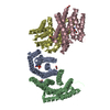
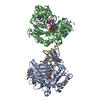
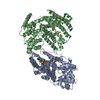
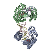
| Title | CRYSTAL STRUCTURE OF THE 14-3-3:NEUTRAL TREHALASE NTH1 COMPLEX | |||||||||
 Components Components |
| |||||||||
 Keywords Keywords | SIGNALING PROTEIN / 14-3-3 / neutral trehalase / hydrolase | |||||||||
| Function / homology |  Function and homology information Function and homology informationChk1/Chk2(Cds1) mediated inactivation of Cyclin B:Cdk1 complex / trehalase activity / RHO GTPases activate PKNs / mitotic spindle orientation checkpoint signaling / signal transduction involved in filamentous growth / alpha,alpha-trehalase / alpha,alpha-trehalase activity / cellular response to desiccation / trehalose catabolic process / pseudohyphal growth ...Chk1/Chk2(Cds1) mediated inactivation of Cyclin B:Cdk1 complex / trehalase activity / RHO GTPases activate PKNs / mitotic spindle orientation checkpoint signaling / signal transduction involved in filamentous growth / alpha,alpha-trehalase / alpha,alpha-trehalase activity / cellular response to desiccation / trehalose catabolic process / pseudohyphal growth / chitin biosynthetic process / regulation of glycogen metabolic process / ascospore formation / HSF1 activation / Regulation of HSF1-mediated heat shock response / aggresome assembly / DNA replication origin binding / phosphoserine residue binding / negative regulation of protein ubiquitination / DNA damage checkpoint signaling / enzyme activator activity / cytoplasmic stress granule / intracellular protein localization / mitotic cell cycle / RNA polymerase II-specific DNA-binding transcription factor binding / Ras protein signal transduction / calcium ion binding / negative regulation of apoptotic process / negative regulation of transcription by RNA polymerase II / signal transduction / nucleus / plasma membrane / cytoplasm Similarity search - Function | |||||||||
| Biological species |  | |||||||||
| Method |  X-RAY DIFFRACTION / SYNCHROTRON / MOLECULAR REPLACEMENT / Resolution: 2.29 Å X-RAY DIFFRACTION / SYNCHROTRON / MOLECULAR REPLACEMENT / Resolution: 2.29 Å | |||||||||
 Authors Authors | Alblova, M. / Smidova, A. / Obsilova, V. / Obsil, T. | |||||||||
 Citation Citation | Journal: Proc. Natl. Acad. Sci. U.S.A. / Year: 2017 Title: Molecular basis of the 14-3-3 protein-dependent activation of yeast neutral trehalase Nth1. Authors: Alblova, M. / Smidova, A. / Docekal, V. / Vesely, J. / Herman, P. / Obsilova, V. / Obsil, T. | |||||||||
| History |
|
- Structure visualization
| Structure viewer | Molecule: MolmilJmol/JSmol |
|---|
- Downloads & links
-Download
| PDBx/mmCIF format | 5n6n.cif.gz | 256.6 KB | Display | PDBx/mmCIF format |
|---|---|---|---|---|
| PDB format | pdb5n6n.ent.gz | 197.9 KB | Display | PDB format |
| PDBx/mmJSON format | 5n6n.json.gz | Tree view | PDBx/mmJSON format | |
| Others |  Other downloads Other downloads |
-Validation report
| Arichive directory | https://data.pdbj.org/pub/pdb/validation_reports/n6/5n6nftp://data.pdbj.org/pub/pdb/validation_reports/n6/5n6n | HTTPS FTP |
|---|
-Related structure data
| Related structure data |  5jtaSC  5m4aC 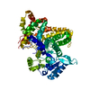 5nisC  2br9S S: Starting model for refinement C: citing same article ( |
|---|---|
| Similar structure data |


-Links
 PDBj
PDBj


- Assembly
| Deposited unit | 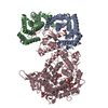
| ||||||||
|---|---|---|---|---|---|---|---|---|---|
| 1 |
| ||||||||
| Unit cell |
|
-Components
| #1: Protein | Mass: 27372.318 Da / Num. of mol.: 2 Source method: isolated from a genetically manipulated source Source: (gene. exp.)  #2: Protein | | Mass: 86670.477 Da / Num. of mol.: 1 Source method: isolated from a genetically manipulated source Source: (gene. exp.) #3: Polysaccharide |   Source method: isolated from a genetically manipulated source Details: oligosaccharide with reducing-end-to-reducing-end glycosidic bond References: sucrose #4: Chemical | ChemComp-CA / |   Mass: 40.078 Da / Num. of mol.: 1 / Source method: obtained synthetically / Formula: Ca Mass: 40.078 Da / Num. of mol.: 1 / Source method: obtained synthetically / Formula: Ca#5: Water | ChemComp-HOH / |  Mass: 18.015 Da / Num. of mol.: 448 / Source method: isolated from a natural source / Formula: H2O Mass: 18.015 Da / Num. of mol.: 448 / Source method: isolated from a natural source / Formula: H2OHas protein modification | Y | |
|---|
-Experimental details
-Experiment
| Experiment | Method: X-RAY DIFFRACTION / Number of used crystals: 1 |
|---|
- Sample preparation
| Crystal | Density Matthews: 2.68 Å3/Da / Density % sol: 54.12 % |
|---|---|
| Crystal grow | Temperature: 291.15 K / Method: vapor diffusion / pH: 6.5 Details: 100 mM sodium cacodylate, 200 mM calcium acetate, 18% (w/v) PEG 8000, 12% (w/v) sucrose |
-Data collection
| Diffraction | Mean temperature: 100 K |
|---|---|
| Diffraction source | Source: SYNCHROTRON / Site: BESSY  / Beamline: 14.1 / Wavelength: 0.9184 Å / Beamline: 14.1 / Wavelength: 0.9184 Å |
| Detector | Type: DECTRIS PILATUS 6M / Detector: PIXEL / Date: Oct 15, 2016 / Details: Sagitally bended Si111-crystal |
| Radiation | Protocol: SINGLE WAVELENGTH / Monochromatic (M) / Laue (L): M / Scattering type: x-ray |
| Radiation wavelength | Wavelength: 0.9184 Å / Relative weight: 1 |
| Reflection | Resolution: 2.29→47.71 Å / Num. obs: 65450 / % possible obs: 97.2 % / Redundancy: 3.5 % / Rsym value: 0.115 / Net I/σ(I): 11.79 |
| Reflection shell | Resolution: 2.29→2.42 Å / Mean I/σ(I) obs: 1.78 / Num. unique obs: 5711 / Rsym value: 0.679 / % possible all: 85.5 |
- Processing
| Software |
| ||||||||||||||||||||||||||||||||||||||||||||||||||||||||||||||||||||||||||||||||||||||||||||||||||||||||||||||||
|---|---|---|---|---|---|---|---|---|---|---|---|---|---|---|---|---|---|---|---|---|---|---|---|---|---|---|---|---|---|---|---|---|---|---|---|---|---|---|---|---|---|---|---|---|---|---|---|---|---|---|---|---|---|---|---|---|---|---|---|---|---|---|---|---|---|---|---|---|---|---|---|---|---|---|---|---|---|---|---|---|---|---|---|---|---|---|---|---|---|---|---|---|---|---|---|---|---|---|---|---|---|---|---|---|---|---|---|---|---|---|---|---|---|
| Refinement | Method to determine structure: MOLECULAR REPLACEMENT Starting model: 5JTA, homology model of Bmh1 based on 2BR9 Resolution: 2.29→46.155 Å / SU ML: 0.28 / Cross valid method: FREE R-VALUE / σ(F): 1.36 / Phase error: 26.02
| ||||||||||||||||||||||||||||||||||||||||||||||||||||||||||||||||||||||||||||||||||||||||||||||||||||||||||||||||
| Solvent computation | Shrinkage radii: 0.9 Å / VDW probe radii: 1.11 Å | ||||||||||||||||||||||||||||||||||||||||||||||||||||||||||||||||||||||||||||||||||||||||||||||||||||||||||||||||
| Refinement step | Cycle: LAST / Resolution: 2.29→46.155 Å
| ||||||||||||||||||||||||||||||||||||||||||||||||||||||||||||||||||||||||||||||||||||||||||||||||||||||||||||||||
| Refine LS restraints |
| ||||||||||||||||||||||||||||||||||||||||||||||||||||||||||||||||||||||||||||||||||||||||||||||||||||||||||||||||
| LS refinement shell |
|
