 Movie
Movie Controller

[English] 日本語
 Yorodumi
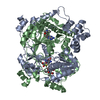
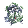
- PDB-5n4i: Crystal structure of OphA-DeltaC6 mutant W400A in complex with SAM -
+ Open data

- Basic information
| Entry | Database: PDB / ID: 5n4i | ||||||
|---|---|---|---|---|---|---|---|
| Title | Crystal structure of OphA-DeltaC6 mutant W400A in complex with SAM | ||||||
 Components Components | Peptide N-methyltransferase | ||||||
 Keywords Keywords | TRANSFERASE / methyltransferase | ||||||
| Function / homology |  Function and homology information Function and homology informationTransferases; Transferring one-carbon groups; Methyltransferases / methyltransferase activity / methylation Similarity search - Function | ||||||
| Biological species |  Omphalotus olearius (fungus) Omphalotus olearius (fungus) | ||||||
| Method |  X-RAY DIFFRACTION / SYNCHROTRON / MOLECULAR REPLACEMENT / Resolution: 1.59 Å X-RAY DIFFRACTION / SYNCHROTRON / MOLECULAR REPLACEMENT / Resolution: 1.59 Å | ||||||
 Authors Authors | Song, H. / Naismith, J.H. | ||||||
 Citation Citation | Journal: Sci Adv / Year: 2018 Title: A molecular mechanism for the enzymatic methylation of nitrogen atoms within peptide bonds. Authors: Song, H. / van der Velden, N.S. / Shiran, S.L. / Bleiziffer, P. / Zach, C. / Sieber, R. / Imani, A.S. / Krausbeck, F. / Aebi, M. / Freeman, M.F. / Riniker, S. / Kunzler, M. / Naismith, J.H. | ||||||
| History |
|
- Structure visualization
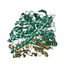
| Structure viewer | Molecule: MolmilJmol/JSmol |
|---|
- Downloads & links
-Download
| PDBx/mmCIF format | 5n4i.cif.gz | 186.3 KB | Display | PDBx/mmCIF format |
|---|---|---|---|---|
| PDB format | pdb5n4i.ent.gz | 147.8 KB | Display | PDB format |
| PDBx/mmJSON format | 5n4i.json.gz | Tree view | PDBx/mmJSON format | |
| Others |  Other downloads Other downloads |
-Validation report
| Arichive directory | https://data.pdbj.org/pub/pdb/validation_reports/n4/5n4iftp://data.pdbj.org/pub/pdb/validation_reports/n4/5n4i | HTTPS FTP |
|---|
-Related structure data
| Related structure data | 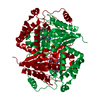 5n0nC 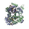 5n0oSC 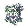 5n0pC  5n0qC 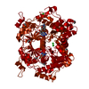 5n0rC 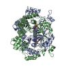 5n0sC 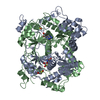 5n0tC 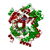 5n0uC 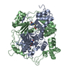 5n0vC 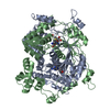 5n0wC 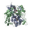 5n0xC 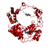 5oufC 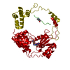 6gewC S: Starting model for refinement C: citing same article ( |
|---|---|
| Similar structure data |


-Links
 PDBj
PDBj


- Assembly
| Deposited unit | 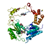
| |||||||||
|---|---|---|---|---|---|---|---|---|---|---|
| 1 | 
| |||||||||
| Unit cell |
| |||||||||
| Components on special symmetry positions |
|
-Components
-Protein , 1 types, 1 molecules A
| #1: Protein | Mass: 44913.059 Da / Num. of mol.: 1 Source method: isolated from a genetically manipulated source Source: (gene. exp.) Omphalotus olearius (fungus) / Production host:  |
|---|
-Non-polymers , 6 types, 243 molecules 


| #2: Chemical | ChemComp-SAM /  Mass: 398.437 Da / Num. of mol.: 1 / Source method: obtained synthetically / Formula: C15H22N6O5S Mass: 398.437 Da / Num. of mol.: 1 / Source method: obtained synthetically / Formula: C15H22N6O5S | ||||||
|---|---|---|---|---|---|---|---|
| #3: Chemical | ChemComp-GOL /  Mass: 92.094 Da / Num. of mol.: 1 / Source method: obtained synthetically / Formula: C3H8O3 Mass: 92.094 Da / Num. of mol.: 1 / Source method: obtained synthetically / Formula: C3H8O3 | ||||||
| #4: Chemical | ChemComp-SCN /  Mass: 58.082 Da / Num. of mol.: 4 / Source method: obtained synthetically / Formula: CNS Mass: 58.082 Da / Num. of mol.: 4 / Source method: obtained synthetically / Formula: CNS#5: Chemical | ChemComp-BCT / |  Mass: 61.017 Da / Num. of mol.: 1 / Source method: obtained synthetically / Formula: CHO3 / Comment: pH buffer*YM Mass: 61.017 Da / Num. of mol.: 1 / Source method: obtained synthetically / Formula: CHO3 / Comment: pH buffer*YM#6: Chemical | ChemComp-MLI / |  Mass: 102.046 Da / Num. of mol.: 1 / Source method: obtained synthetically / Formula: C3H2O4 Mass: 102.046 Da / Num. of mol.: 1 / Source method: obtained synthetically / Formula: C3H2O4#7: Water | ChemComp-HOH / | Mass: 18.015 Da / Num. of mol.: 235 / Source method: isolated from a natural source / Formula: H2O |
-Details
| Has protein modification | Y |
|---|
-Experimental details
-Experiment
| Experiment | Method: X-RAY DIFFRACTION / Number of used crystals: 1 |
|---|
- Sample preparation
| Crystal | Density Matthews: 3.46 Å3/Da / Density % sol: 64.42 % |
|---|---|
| Crystal grow | Temperature: 289 K / Method: vapor diffusion, hanging drop Details: 0.3-0.4 M KSCN, 0.1 M Bicine pH 9.0, 1.7-1.9 M sodium malonate |
-Data collection
| Diffraction | Mean temperature: 100 K |
|---|---|
| Diffraction source | Source: SYNCHROTRON / Site: Diamond  / Beamline: I03 / Wavelength: 0.9763 Å / Beamline: I03 / Wavelength: 0.9763 Å |
| Detector | Type: DECTRIS PILATUS3 6M / Detector: PIXEL / Date: Jan 21, 2017 |
| Radiation | Protocol: SINGLE WAVELENGTH / Monochromatic (M) / Laue (L): M / Scattering type: x-ray |
| Radiation wavelength | Wavelength: 0.9763 Å / Relative weight: 1 |
| Reflection | Resolution: 1.59→75.9 Å / Num. obs: 85158 / % possible obs: 98.6 % / Redundancy: 7.3 % / CC1/2: 0.997 / Rmerge(I) obs: 0.045 / Net I/σ(I): 14.9 |
| Reflection shell | Resolution: 1.59→1.62 Å / Redundancy: 7.2 % / Rmerge(I) obs: 1.545 / Mean I/σ(I) obs: 1.2 / CC1/2: 0.605 / % possible all: 95.3 |
- Processing
| Software |
| ||||||||||||||||||||||||||||||||||||||||||||||||||||||||||||||||||||||||||||||||||||||||||||||||||||||||||||||||||||||||||||||||||||||||||||||||||||||||||||||||||||||||||||||||||||||
|---|---|---|---|---|---|---|---|---|---|---|---|---|---|---|---|---|---|---|---|---|---|---|---|---|---|---|---|---|---|---|---|---|---|---|---|---|---|---|---|---|---|---|---|---|---|---|---|---|---|---|---|---|---|---|---|---|---|---|---|---|---|---|---|---|---|---|---|---|---|---|---|---|---|---|---|---|---|---|---|---|---|---|---|---|---|---|---|---|---|---|---|---|---|---|---|---|---|---|---|---|---|---|---|---|---|---|---|---|---|---|---|---|---|---|---|---|---|---|---|---|---|---|---|---|---|---|---|---|---|---|---|---|---|---|---|---|---|---|---|---|---|---|---|---|---|---|---|---|---|---|---|---|---|---|---|---|---|---|---|---|---|---|---|---|---|---|---|---|---|---|---|---|---|---|---|---|---|---|---|---|---|---|---|
| Refinement | Method to determine structure: MOLECULAR REPLACEMENT Starting model: 5N0O Resolution: 1.59→75.9 Å / Cor.coef. Fo:Fc: 0.976 / Cor.coef. Fo:Fc free: 0.969 / SU B: 4.699 / SU ML: 0.066 / Cross valid method: THROUGHOUT / ESU R: 0.075 / ESU R Free: 0.069 / Stereochemistry target values: MAXIMUM LIKELIHOOD / Details: HYDROGENS HAVE BEEN ADDED IN THE RIDING POSITIONS
| ||||||||||||||||||||||||||||||||||||||||||||||||||||||||||||||||||||||||||||||||||||||||||||||||||||||||||||||||||||||||||||||||||||||||||||||||||||||||||||||||||||||||||||||||||||||
| Solvent computation | Ion probe radii: 0.8 Å / Shrinkage radii: 0.8 Å / VDW probe radii: 1.2 Å / Solvent model: MASK | ||||||||||||||||||||||||||||||||||||||||||||||||||||||||||||||||||||||||||||||||||||||||||||||||||||||||||||||||||||||||||||||||||||||||||||||||||||||||||||||||||||||||||||||||||||||
| Displacement parameters | Biso mean: 41.61 Å2
| ||||||||||||||||||||||||||||||||||||||||||||||||||||||||||||||||||||||||||||||||||||||||||||||||||||||||||||||||||||||||||||||||||||||||||||||||||||||||||||||||||||||||||||||||||||||
| Refinement step | Cycle: 1 / Resolution: 1.59→75.9 Å
| ||||||||||||||||||||||||||||||||||||||||||||||||||||||||||||||||||||||||||||||||||||||||||||||||||||||||||||||||||||||||||||||||||||||||||||||||||||||||||||||||||||||||||||||||||||||
| Refine LS restraints |
|