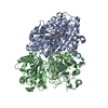
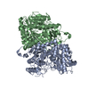
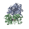
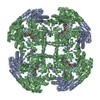
Mass: 63091.121 Da / Num. of mol.: 2 Source method: isolated from a genetically manipulated source Details: The residues that are not present in the coordinates are disordered and not visible in the electron density. They are not included in the final model. Side chain atoms of residues His149A, ...Details: The residues that are not present in the coordinates are disordered and not visible in the electron density. They are not included in the final model. Side chain atoms of residues His149A, Ser328A, His149B, Ser328B which lie on the protein surface" have been included in the pdb but set at zero occupancy because they completely lack electron density. Source: (gene. exp.) Penicillium simplicissimum (fungus) / Gene: VAOA / Production host: Escherichia coli (E. coli) / References: UniProt: P56216, vanillyl-alcohol oxidase
Resolution: 2.8→54.94 Å / Cor.coef. Fo:Fc: 0.954 / Cor.coef. Fo:Fc free: 0.901 / SU B: 18.61 / SU ML: 0.34 / Cross valid method: THROUGHOUT / ESU R Free: 0.428 / Stereochemistry target values: MAXIMUM LIKELIHOOD / Details: HYDROGENS HAVE BEEN ADDED IN THE RIDING POSITIONS
Rfactor
Num. reflection
% reflection
Selection details
Rfree
0.24886
1353
4.9 %
RANDOM
Rwork
0.17522
-
-
-
obs
0.17884
26493
90.69 %
-
Solvent computation
Ion probe radii: 0.8 Å / Shrinkage radii: 0.8 Å / VDW probe radii: 1.2 Å / Solvent model: MASK
 Movie
Movie Controller


 Open data
 Basic information
 Components
 Keywords
 Function and homology information
 Penicillium simplicissimum (fungus)
 X-RAY DIFFRACTION /
  Authors
 Netherlands,
  Italy, 2items
  Citation
 Structure visualization
 Downloads & links
 Other downloads


 PDBj
PDBj
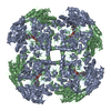
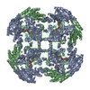
 Assembly


 Mass: 785.550 Da / Num. of mol.: 2 / Source method: obtained synthetically / Formula: C27H33N9O15P2 / Comment: FAD*YM
 Mass: 92.094 Da / Num. of mol.: 2 / Source method: obtained synthetically / Formula: C3H8O3
 Mass: 18.015 Da / Num. of mol.: 232 / Source method: isolated from a natural source / Formula: H2O
 Sample preparation
 / Beamline: X06DA / Wavelength: 0.999 / Wavelength: 0.999 Å
 Processing