 Movie
Movie Controller

[English] 日本語
 Yorodumi
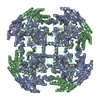
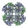
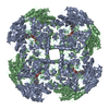
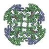
- PDB-1e8f: STRUCTURE OF THE H61T MUTANT OF THE FLAVOENZYME VANILLYL-ALCOHOL ... -
+ Open data

- Basic information
| Entry | Database: PDB / ID: 1e8f | ||||||
|---|---|---|---|---|---|---|---|
| Title | STRUCTURE OF THE H61T MUTANT OF THE FLAVOENZYME VANILLYL-ALCOHOL OXIDASE IN THE APO FORM | ||||||
 Components Components | VANILLYL-ALCOHOL OXIDASE | ||||||
 Keywords Keywords | OXIDOREDUCTASE / FLAVOPROTEIN / METHANOL UTILIZATION / PEROXISOME / FLAVOENZYME / OXIDASE / CATALYSIS | ||||||
| Function / homology |  Function and homology information Function and homology informationvanillyl-alcohol oxidase / vanillyl-alcohol oxidase activity / methanol metabolic process / lactate catabolic process / D-lactate dehydrogenase (cytochrome) activity / D-lactate dehydrogenase (NAD+) activity / FAD binding / peroxisome / mitochondrion Similarity search - Function | ||||||
| Biological species |  PENICILLIUM SIMPLICISSIMUM (fungus) PENICILLIUM SIMPLICISSIMUM (fungus) | ||||||
| Method |  X-RAY DIFFRACTION / SYNCHROTRON / MOLECULAR REPLACEMENT / Resolution: 2.9 Å X-RAY DIFFRACTION / SYNCHROTRON / MOLECULAR REPLACEMENT / Resolution: 2.9 Å | ||||||
 Authors Authors | Mattevi, A. / Fraaije, M.W. | ||||||
 Citation Citation | Journal: J.Biol.Chem. / Year: 2001 Title: Structural Analysis of Flavinylation in Vanillyl-Alcohol Oxidase Authors: Fraaije, M.W. / Van Der Heuvel, R.H.H. / Van Berkel, W.J.H. / Mattevi, A. | ||||||
| History |
|
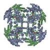
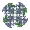
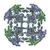
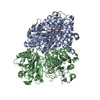
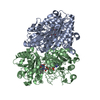
- Structure visualization
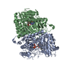
| Structure viewer | Molecule: MolmilJmol/JSmol |
|---|
- Downloads & links
-Download
| PDBx/mmCIF format | 1e8f.cif.gz | 222.7 KB | Display | PDBx/mmCIF format |
|---|---|---|---|---|
| PDB format | pdb1e8f.ent.gz | 176.7 KB | Display | PDB format |
| PDBx/mmJSON format | 1e8f.json.gz | Tree view | PDBx/mmJSON format | |
| Others |  Other downloads Other downloads |
-Validation report
| Arichive directory | https://data.pdbj.org/pub/pdb/validation_reports/e8/1e8fftp://data.pdbj.org/pub/pdb/validation_reports/e8/1e8f | HTTPS FTP |
|---|
-Related structure data
| Related structure data |  1e8gC  1e8hC  1qltS C: citing same article ( S: Starting model for refinement |
|---|---|
| Similar structure data |


-Links
 PDBj
PDBj
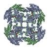
- Assembly
| Deposited unit | 
| ||||||||
|---|---|---|---|---|---|---|---|---|---|
| 1 | 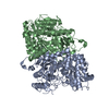
| ||||||||
| Unit cell |
| ||||||||
| Noncrystallographic symmetry (NCS) | NCS oper: (Code: given Matrix: (0.236096, -0.97158, 0.017062), Vector: |
-Components
| #1: Protein | Mass: 62969.934 Da / Num. of mol.: 2 / Mutation: YES Source method: isolated from a genetically manipulated source Source: (gene. exp.) PENICILLIUM SIMPLICISSIMUM (fungus) / Description: FUNGUS / Cellular location: INTRACELLULAR / Organelle: PEROXISOMES / Production host:  #2: Water | ChemComp-HOH / |  Mass: 18.015 Da / Num. of mol.: 20 / Source method: isolated from a natural source / Formula: H2O Mass: 18.015 Da / Num. of mol.: 20 / Source method: isolated from a natural source / Formula: H2OCompound details | CHAIN A, B ENGINEERED MUTATION HIS61THR CATALYZES THE CONVERSION OF VANILLIN ALCOHOL TO VANILLIN, ...CHAIN A, B ENGINEERED | |
|---|
-Experimental details
-Experiment
| Experiment | Method: X-RAY DIFFRACTION / Number of used crystals: 1 |
|---|
- Sample preparation
| Crystal | Density Matthews: 2.3 Å3/Da / Density % sol: 43.14 % | |||||||||||||||
|---|---|---|---|---|---|---|---|---|---|---|---|---|---|---|---|---|
| Crystal grow | pH: 4.6 / Details: FROM 100 MM ACETATE BUFFER PH 4.6, 6% PEG4000 | |||||||||||||||
| Crystal grow | *PLUS pH: 5.1 / Method: vapor diffusion, hanging drop | |||||||||||||||
| Components of the solutions | *PLUS
|
-Data collection
| Diffraction | Mean temperature: 100 K |
|---|---|
| Diffraction source | Source: SYNCHROTRON / Site: ESRF  / Beamline: ID14-3 / Wavelength: 1 / Beamline: ID14-3 / Wavelength: 1 |
| Detector | Type: MARRESEARCH / Detector: IMAGE PLATE / Date: Feb 15, 1999 / Details: MIRRORS |
| Radiation | Monochromator: NI FILTER / Protocol: SINGLE WAVELENGTH / Monochromatic (M) / Laue (L): M / Scattering type: x-ray |
| Radiation wavelength | Wavelength: 1 Å / Relative weight: 1 |
| Reflection | Resolution: 2.9→20 Å / Num. obs: 23727 / % possible obs: 98.1 % / Observed criterion σ(I): 0 / Redundancy: 2.3 % / Rmerge(I) obs: 0.135 / Rsym value: 0.135 / Net I/σ(I): 6 |
| Reflection shell | Resolution: 2.8→2.9 Å / Redundancy: 2.2 % / Rmerge(I) obs: 0.493 / Mean I/σ(I) obs: 2.8 / Rsym value: 0.493 / % possible all: 98.1 |
| Reflection | *PLUS Num. measured all: 54479 |
| Reflection shell | *PLUS % possible obs: 98.1 % |
- Processing
| Software |
| ||||||||||||||||||||||||||||||||||||||||||||||||||||||||||||||||||||||||||||||||||||
|---|---|---|---|---|---|---|---|---|---|---|---|---|---|---|---|---|---|---|---|---|---|---|---|---|---|---|---|---|---|---|---|---|---|---|---|---|---|---|---|---|---|---|---|---|---|---|---|---|---|---|---|---|---|---|---|---|---|---|---|---|---|---|---|---|---|---|---|---|---|---|---|---|---|---|---|---|---|---|---|---|---|---|---|---|---|
| Refinement | Method to determine structure: MOLECULAR REPLACEMENT Starting model: 1QLT Resolution: 2.9→20 Å / Cross valid method: THROUGHOUT / σ(F): 0
| ||||||||||||||||||||||||||||||||||||||||||||||||||||||||||||||||||||||||||||||||||||
| Refinement step | Cycle: LAST / Resolution: 2.9→20 Å
| ||||||||||||||||||||||||||||||||||||||||||||||||||||||||||||||||||||||||||||||||||||
| Refine LS restraints |
| ||||||||||||||||||||||||||||||||||||||||||||||||||||||||||||||||||||||||||||||||||||
| Software | *PLUS Name: REFMAC / Classification: refinement | ||||||||||||||||||||||||||||||||||||||||||||||||||||||||||||||||||||||||||||||||||||
| Refine LS restraints | *PLUS Type: p_plane_restr / Dev ideal: 0.011 |
