 Movie
Movie Controller


+ Open data

- Basic information
| Entry | Database: PDB / ID: 5mum | ||||||
|---|---|---|---|---|---|---|---|
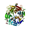
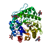
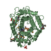
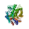
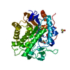
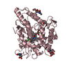
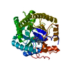
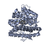
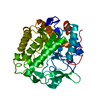
| Title | Glycoside Hydrolase BACINT_00347 | ||||||
 Components Components | BACINT_00347 | ||||||
 Keywords Keywords | HYDROLASE / Rhamnosidase / Bacteroides / Beta-propeller | ||||||
| Function / homology | BNR repeat-containing family member / Sialidase superfamily / Neuraminidase Function and homology information Function and homology information | ||||||
| Biological species |  Bacteroides intestinalis DSM 17393 (bacteria) Bacteroides intestinalis DSM 17393 (bacteria) | ||||||
| Method |  X-RAY DIFFRACTION / SYNCHROTRON / MOLECULAR REPLACEMENT / Resolution: 1.8 Å X-RAY DIFFRACTION / SYNCHROTRON / MOLECULAR REPLACEMENT / Resolution: 1.8 Å | ||||||
 Authors Authors | Munoz-Munoz, J. / Cartmell, A. / Terrapon, N. / Henrissat, B. / Gilbert, H.J. | ||||||
| Funding support |  United Kingdom, 1items United Kingdom, 1items
| ||||||
 Citation Citation | Journal: Proc. Natl. Acad. Sci. U.S.A. / Year: 2017 Title: Unusual active site location and catalytic apparatus in a glycoside hydrolase family. Authors: Munoz-Munoz, J. / Cartmell, A. / Terrapon, N. / Henrissat, B. / Gilbert, H.J. | ||||||
| History |
|
- Structure visualization
| Structure viewer | Molecule: MolmilJmol/JSmol |
|---|
- Downloads & links
-Download
| PDBx/mmCIF format | 5mum.cif.gz | 103.2 KB | Display | PDBx/mmCIF format |
|---|---|---|---|---|
| PDB format | pdb5mum.ent.gz | 77.3 KB | Display | PDB format |
| PDBx/mmJSON format | 5mum.json.gz | Tree view | PDBx/mmJSON format | |
| Others |  Other downloads Other downloads |
-Validation report
| Arichive directory | https://data.pdbj.org/pub/pdb/validation_reports/mu/5mumftp://data.pdbj.org/pub/pdb/validation_reports/mu/5mum | HTTPS FTP |
|---|
-Related structure data
| Related structure data | 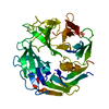 5mukC 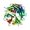 5mulC 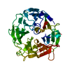 5mvhC  4irtS S: Starting model for refinement C: citing same article ( |
|---|---|
| Similar structure data |

-Links
 PDBj
PDBj

- Assembly
| Deposited unit | 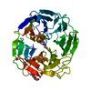
| ||||||||
|---|---|---|---|---|---|---|---|---|---|
| 1 |
| ||||||||
| Unit cell |
|
-Components
| #1: Protein | Mass: 49838.141 Da / Num. of mol.: 1 Source method: isolated from a genetically manipulated source Source: (gene. exp.) Bacteroides intestinalis DSM 17393 (bacteria)Gene: BACINT_00347 / Production host: | ||||
|---|---|---|---|---|---|
| #2: Chemical |   Mass: 62.068 Da / Num. of mol.: 2 / Source method: obtained synthetically / Formula: C2H6O2 Mass: 62.068 Da / Num. of mol.: 2 / Source method: obtained synthetically / Formula: C2H6O2#3: Chemical |   Mass: 238.278 Da / Num. of mol.: 2 / Source method: obtained synthetically / Formula: C10H22O6 / Comment: precipitant*YM Mass: 238.278 Da / Num. of mol.: 2 / Source method: obtained synthetically / Formula: C10H22O6 / Comment: precipitant*YM#4: Water | ChemComp-HOH / |  Mass: 18.015 Da / Num. of mol.: 314 / Source method: isolated from a natural source / Formula: H2O Mass: 18.015 Da / Num. of mol.: 314 / Source method: isolated from a natural source / Formula: H2O |
-Experimental details
-Experiment
| Experiment | Method: X-RAY DIFFRACTION / Number of used crystals: 1 |
|---|
- Sample preparation
| Crystal | Density Matthews: 2.17 Å3/Da / Density % sol: 43.38 % |
|---|---|
| Crystal grow | Temperature: 293 K / Method: vapor diffusion, sitting drop / Details: 20% PEG3350 and 200 mM Tri-potassium Citrate |
-Data collection
| Diffraction | Mean temperature: 100 K |
|---|---|
| Diffraction source | Source: SYNCHROTRON / Site: Diamond / Beamline: I03 / Wavelength: 0.979 Å |
| Detector | Type: DECTRIS PILATUS3 6M / Detector: PIXEL / Date: May 15, 2015 |
| Radiation | Protocol: SINGLE WAVELENGTH / Monochromatic (M) / Laue (L): M / Scattering type: x-ray |
| Radiation wavelength | Wavelength: 0.979 Å / Relative weight: 1 |
| Reflection | Resolution: 1.8→68.6 Å / Num. obs: 255841 / % possible obs: 98.4 % / Redundancy: 3.3 % / Net I/σ(I): 12 |
- Processing
| Software |
| ||||||||||||||||||||||||||||||||||||||||||||||||||||||||||||||||||||||||||||||||||||||||||||||||||||||||||||||||||||||||||||||||||||||||||||||||||||||||||||||||||||||||||||||||||||||
|---|---|---|---|---|---|---|---|---|---|---|---|---|---|---|---|---|---|---|---|---|---|---|---|---|---|---|---|---|---|---|---|---|---|---|---|---|---|---|---|---|---|---|---|---|---|---|---|---|---|---|---|---|---|---|---|---|---|---|---|---|---|---|---|---|---|---|---|---|---|---|---|---|---|---|---|---|---|---|---|---|---|---|---|---|---|---|---|---|---|---|---|---|---|---|---|---|---|---|---|---|---|---|---|---|---|---|---|---|---|---|---|---|---|---|---|---|---|---|---|---|---|---|---|---|---|---|---|---|---|---|---|---|---|---|---|---|---|---|---|---|---|---|---|---|---|---|---|---|---|---|---|---|---|---|---|---|---|---|---|---|---|---|---|---|---|---|---|---|---|---|---|---|---|---|---|---|---|---|---|---|---|---|---|
| Refinement | Method to determine structure: MOLECULAR REPLACEMENT Starting model: 4IRT Resolution: 1.8→68.6 Å / Cor.coef. Fo:Fc: 0.957 / Cor.coef. Fo:Fc free: 0.932 / SU B: 2.495 / SU ML: 0.075 / Cross valid method: THROUGHOUT / ESU R: 0.127 / ESU R Free: 0.121 / Details: HYDROGENS HAVE BEEN ADDED IN THE RIDING POSITIONS
| ||||||||||||||||||||||||||||||||||||||||||||||||||||||||||||||||||||||||||||||||||||||||||||||||||||||||||||||||||||||||||||||||||||||||||||||||||||||||||||||||||||||||||||||||||||||
| Solvent computation | Ion probe radii: 0.8 Å / Shrinkage radii: 0.8 Å / VDW probe radii: 1.2 Å | ||||||||||||||||||||||||||||||||||||||||||||||||||||||||||||||||||||||||||||||||||||||||||||||||||||||||||||||||||||||||||||||||||||||||||||||||||||||||||||||||||||||||||||||||||||||
| Displacement parameters | Biso mean: 13.636 Å2
| ||||||||||||||||||||||||||||||||||||||||||||||||||||||||||||||||||||||||||||||||||||||||||||||||||||||||||||||||||||||||||||||||||||||||||||||||||||||||||||||||||||||||||||||||||||||
| Refinement step | Cycle: 1 / Resolution: 1.8→68.6 Å
| ||||||||||||||||||||||||||||||||||||||||||||||||||||||||||||||||||||||||||||||||||||||||||||||||||||||||||||||||||||||||||||||||||||||||||||||||||||||||||||||||||||||||||||||||||||||
| Refine LS restraints |
|