 Movie
Movie Controller

[English] 日本語
 Yorodumi
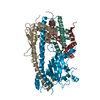
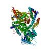
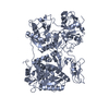
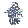
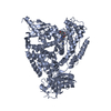
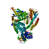
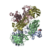
- PDB-5mu4: Tail Tubular Protein A of Klebsiella pneumoniae bacteriophage KP32 -
+ Open data

- Basic information
| Entry | Database: PDB / ID: 5mu4 | ||||||
|---|---|---|---|---|---|---|---|
| Title | Tail Tubular Protein A of Klebsiella pneumoniae bacteriophage KP32 | ||||||
 Components Components | Tail tubular protein A | ||||||
 Keywords Keywords | VIRAL PROTEIN / tail tubular protein / bacteriophage / exopolysaccharide depolymerase / antibacterial activity | ||||||
| Function / homology | Tail tubular protein Gp11 / Tail tubular protein / symbiont entry into host cell via disruption of host cell glycocalyx / symbiont entry into host cell via disruption of host cell envelope / hydrolase activity, acting on glycosyl bonds / Hydrolases; Glycosylases; Glycosidases, i.e. enzymes that hydrolyse O- and S-glycosyl compounds / adhesion receptor-mediated virion attachment to host cell / virion component / Tail tubular protein A Function and homology information Function and homology information | ||||||
| Biological species |  Klebsiella phage KP32 (virus) Klebsiella phage KP32 (virus) | ||||||
| Method |  X-RAY DIFFRACTION / SYNCHROTRON / SAD / Resolution: 1.9 Å X-RAY DIFFRACTION / SYNCHROTRON / SAD / Resolution: 1.9 Å | ||||||
 Authors Authors | Pyra, A. / Brzozowska, E. / Pawlik, K. / Dauter, M. / Dauter, Z. / Gamian, A. | ||||||
 Citation Citation | Journal: Sci Rep / Year: 2017 Title: Tail tubular protein A: a dual-function tail protein of Klebsiella pneumoniae bacteriophage KP32. Authors: Pyra, A. / Brzozowska, E. / Pawlik, K. / Gamian, A. / Dauter, M. / Dauter, Z. | ||||||
| History |
|
- Structure visualization
| Structure viewer | Molecule: MolmilJmol/JSmol |
|---|
- Downloads & links
-Download
| PDBx/mmCIF format | 5mu4.cif.gz | 173.6 KB | Display | PDBx/mmCIF format |
|---|---|---|---|---|
| PDB format | pdb5mu4.ent.gz | 138.9 KB | Display | PDB format |
| PDBx/mmJSON format | 5mu4.json.gz | Tree view | PDBx/mmJSON format | |
| Others |  Other downloads Other downloads |
-Validation report
| Arichive directory | https://data.pdbj.org/pub/pdb/validation_reports/mu/5mu4ftp://data.pdbj.org/pub/pdb/validation_reports/mu/5mu4 | HTTPS FTP |
|---|
-Related structure data
| Similar structure data |
|---|


-Links
 PDBj
PDBj- Assembly
| Deposited unit | 
| ||||||||
|---|---|---|---|---|---|---|---|---|---|
| 1 |
| ||||||||
| Unit cell |
|
-Components
| #1: Protein | Mass: 22281.680 Da / Num. of mol.: 4 Source method: isolated from a genetically manipulated source Source: (gene. exp.) Klebsiella phage KP32 (virus) / Gene: 31Production host:  References: UniProt: D1L2W4 #2: Water | ChemComp-HOH / |  Mass: 18.015 Da / Num. of mol.: 791 / Source method: isolated from a natural source / Formula: H2O Mass: 18.015 Da / Num. of mol.: 791 / Source method: isolated from a natural source / Formula: H2O |
|---|
-Experimental details
-Experiment
| Experiment | Method: X-RAY DIFFRACTION / Number of used crystals: 1 |
|---|
- Sample preparation
| Crystal | Density Matthews: 3.19 Å3/Da / Density % sol: 61.43 % |
|---|---|
| Crystal grow | Temperature: 293 K / Method: vapor diffusion / Details: 35% Tacsimate |
-Data collection
| Diffraction |
| |||||||||||||||||||||||||||||||||||||||||||||||||||||||||||||||||||||||||||||||||||||||||||||||||||||||||||||||||||||||||||||||||||||||||||||||||||||||||||||||||||||||||||||||||||||||||||||
|---|---|---|---|---|---|---|---|---|---|---|---|---|---|---|---|---|---|---|---|---|---|---|---|---|---|---|---|---|---|---|---|---|---|---|---|---|---|---|---|---|---|---|---|---|---|---|---|---|---|---|---|---|---|---|---|---|---|---|---|---|---|---|---|---|---|---|---|---|---|---|---|---|---|---|---|---|---|---|---|---|---|---|---|---|---|---|---|---|---|---|---|---|---|---|---|---|---|---|---|---|---|---|---|---|---|---|---|---|---|---|---|---|---|---|---|---|---|---|---|---|---|---|---|---|---|---|---|---|---|---|---|---|---|---|---|---|---|---|---|---|---|---|---|---|---|---|---|---|---|---|---|---|---|---|---|---|---|---|---|---|---|---|---|---|---|---|---|---|---|---|---|---|---|---|---|---|---|---|---|---|---|---|---|---|---|---|---|---|---|---|
| Diffraction source |
| |||||||||||||||||||||||||||||||||||||||||||||||||||||||||||||||||||||||||||||||||||||||||||||||||||||||||||||||||||||||||||||||||||||||||||||||||||||||||||||||||||||||||||||||||||||||||||||
| Detector |
| |||||||||||||||||||||||||||||||||||||||||||||||||||||||||||||||||||||||||||||||||||||||||||||||||||||||||||||||||||||||||||||||||||||||||||||||||||||||||||||||||||||||||||||||||||||||||||||
| Radiation |
| |||||||||||||||||||||||||||||||||||||||||||||||||||||||||||||||||||||||||||||||||||||||||||||||||||||||||||||||||||||||||||||||||||||||||||||||||||||||||||||||||||||||||||||||||||||||||||||
| Radiation wavelength |
| |||||||||||||||||||||||||||||||||||||||||||||||||||||||||||||||||||||||||||||||||||||||||||||||||||||||||||||||||||||||||||||||||||||||||||||||||||||||||||||||||||||||||||||||||||||||||||||
| Reflection | Resolution: 1.9→30 Å / Num. obs: 87540 / % possible obs: 100 % / Redundancy: 8.2 % / Rmerge(I) obs: 0.107 / Rpim(I) all: 0.04 / Rrim(I) all: 0.113 / Χ2: 0.933 / Net I/σ(I): 9.4 / Num. measured all: 718685 | |||||||||||||||||||||||||||||||||||||||||||||||||||||||||||||||||||||||||||||||||||||||||||||||||||||||||||||||||||||||||||||||||||||||||||||||||||||||||||||||||||||||||||||||||||||||||||||
| Reflection shell | Diffraction-ID: 1
|

- Processing
| Software |
| ||||||||||||||||||||||||||||||||||||||||||||||||||||||||||||
|---|---|---|---|---|---|---|---|---|---|---|---|---|---|---|---|---|---|---|---|---|---|---|---|---|---|---|---|---|---|---|---|---|---|---|---|---|---|---|---|---|---|---|---|---|---|---|---|---|---|---|---|---|---|---|---|---|---|---|---|---|---|
| Refinement | Method to determine structure: SAD / Resolution: 1.9→29.95 Å / Cor.coef. Fo:Fc: 0.951 / Cor.coef. Fo:Fc free: 0.925 / SU B: 3.403 / SU ML: 0.098 / Cross valid method: THROUGHOUT / σ(F): 0 / ESU R: 0.124 / ESU R Free: 0.128 / Stereochemistry target values: MAXIMUM LIKELIHOOD Details: HYDROGENS HAVE BEEN ADDED IN THE RIDING POSITIONS U VALUES : REFINED INDIVIDUALLY
| ||||||||||||||||||||||||||||||||||||||||||||||||||||||||||||
| Solvent computation | Ion probe radii: 0.8 Å / Shrinkage radii: 0.8 Å / VDW probe radii: 1.2 Å / Solvent model: BABINET MODEL WITH MASK | ||||||||||||||||||||||||||||||||||||||||||||||||||||||||||||
| Displacement parameters | Biso max: 112.67 Å2 / Biso mean: 28.628 Å2 / Biso min: 8.72 Å2
| ||||||||||||||||||||||||||||||||||||||||||||||||||||||||||||
| Refinement step | Cycle: final / Resolution: 1.9→29.95 Å
| ||||||||||||||||||||||||||||||||||||||||||||||||||||||||||||
| Refine LS restraints |
| ||||||||||||||||||||||||||||||||||||||||||||||||||||||||||||
| LS refinement shell | Resolution: 1.903→1.953 Å / Rfactor Rfree error: 0 / Total num. of bins used: 20
|
