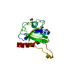
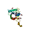
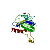
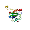
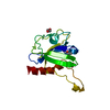
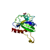
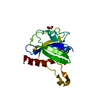
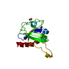
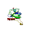
Entry Database : PDB / ID : 5mp0Title Human m7GpppN-mRNA Hydrolase (DCP2, NUDT20) Catalytic Domain m7GpppN-mRNA hydrolase Keywords / / Function / homology Function Domain/homology Component
/ / / / / / / / / / / / / / / / / / / / / / / / / / / / / / / / / / / / / / / / / / / / / / / / / / / / / / / / / Biological species Homo sapiens (human)Method / / / Resolution : 1.63 Å Authors Mathea, S. / Salah, E. / Velupillai, S. / Tallant, C. / Pike, A.C.W. / Bushell, S.R. / Faust, B. / Wang, D. / Burgess-Brown, N. / von Delft, F. ...Mathea, S. / Salah, E. / Velupillai, S. / Tallant, C. / Pike, A.C.W. / Bushell, S.R. / Faust, B. / Wang, D. / Burgess-Brown, N. / von Delft, F. / Arrowsmith, C.H. / Edwards, A.M. / Bountra, C. / Knapp, S. / Huber, K. Journal : To Be Published Title : Human m7GpppN-mRNA Hydrolase (DCP2, NUDT20) Catalytic DomainAuthors : Mathea, S. / Huber, K. History Deposition Dec 15, 2016 Deposition site / Processing site Revision 1.0 Jun 28, 2017 Provider / Type Revision 1.1 Jan 17, 2024 Group / Database references / Refinement descriptionCategory chem_comp_atom / chem_comp_bond ... chem_comp_atom / chem_comp_bond / database_2 / pdbx_initial_refinement_model Item / _database_2.pdbx_database_accession
Show all Show less
 Movie
Movie Controller


 Open data
 Basic information
 Components
 Keywords
 Function and homology information
 Homo sapiens (human)
 X-RAY DIFFRACTION /
  Authors
 Citation
 Structure visualization
 Downloads & links
 Other downloads
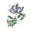

 PDBj
PDBj


 Assembly


 Mass: 62.068 Da / Num. of mol.: 2 / Source method: obtained synthetically / Formula: C2H6O2
 Mass: 18.015 Da / Num. of mol.: 60 / Source method: isolated from a natural source / Formula: H2O
 Sample preparation
 / Beamline: I24 / Wavelength: 0.9686 Å
 Processing