 Movie
Movie Controller


+ Open data

- Basic information
| Entry | Database: PDB / ID: 5mm2 | ||||||||||||
|---|---|---|---|---|---|---|---|---|---|---|---|---|---|
| Title | nora virus structure | ||||||||||||
 Components Components |
| ||||||||||||
 Keywords Keywords | VIRUS / nora virus / cryo-em / single particle analysis / capsid | ||||||||||||
| Function / homology | viral capsid / Capsid protein / ORF4 polyprotein Function and homology information Function and homology information | ||||||||||||
| Biological species |  Nora virus Nora virus | ||||||||||||
| Method | ELECTRON MICROSCOPY / single particle reconstruction / cryo EM / Resolution: 2.7 Å | ||||||||||||
 Authors Authors | Laurinmaki, P. / Shakeel, S. / Ekstrom, J.-O. / Butcher, S.J. | ||||||||||||
| Funding support |  Finland, 3items Finland, 3items
| ||||||||||||
 Citation Citation | Journal: Sci Rep / Year: 2020 Title: Structure of Nora virus at 2.7 Å resolution and implications for receptor binding, capsid stability and taxonomy. Authors: Pasi Laurinmäki / Shabih Shakeel / Jens-Ola Ekström / Pezhman Mohammadi / Dan Hultmark / Sarah J Butcher /   Abstract: Nora virus, a virus of Drosophila, encapsidates one of the largest single-stranded RNA virus genomes known. Its taxonomic affinity is uncertain as it has a picornavirus-like cassette of enzymes for ...Nora virus, a virus of Drosophila, encapsidates one of the largest single-stranded RNA virus genomes known. Its taxonomic affinity is uncertain as it has a picornavirus-like cassette of enzymes for virus replication, but the capsid structure was at the time for genome publication unknown. By solving the structure of the virus, and through sequence comparison, we clear up this taxonomic ambiguity in the invertebrate RNA virosphere. Despite the lack of detectable similarity in the amino acid sequences, the 2.7 Å resolution cryoEM map showed Nora virus to have T = 1 symmetry with the characteristic capsid protein β-barrels found in all the viruses in the Picornavirales order. Strikingly, α-helical bundles formed from the extended C-termini of capsid protein VP4B and VP4C protrude from the capsid surface. They are similar to signalling molecule folds and implicated in virus entry. Unlike other viruses of Picornavirales, no intra-pentamer stabilizing annulus was seen, instead the intra-pentamer stability comes from the interaction of VP4C and VP4B N-termini. Finally, intertwining of the N-termini of two-fold symmetry-related VP4A capsid proteins and RNA, provides inter-pentamer stability. Based on its distinct structural elements and the genetic distance to other picorna-like viruses we propose that Nora virus, and a small group of related viruses, should have its own family within the order Picornavirales. | ||||||||||||
| History |
|
- Structure visualization
| Movie |
Movie viewer |
|---|---|
| Structure viewer | Molecule: MolmilJmol/JSmol |
- Downloads & links
-Download
| PDBx/mmCIF format | 5mm2.cif.gz | 157.3 KB | Display | PDBx/mmCIF format |
|---|---|---|---|---|
| PDB format | pdb5mm2.ent.gz | 118.4 KB | Display | PDB format |
| PDBx/mmJSON format | 5mm2.json.gz | Tree view | PDBx/mmJSON format | |
| Others |  Other downloads Other downloads |
-Validation report
| Arichive directory | https://data.pdbj.org/pub/pdb/validation_reports/mm/5mm2ftp://data.pdbj.org/pub/pdb/validation_reports/mm/5mm2 | HTTPS FTP |
|---|
-Related structure data
| Related structure data | 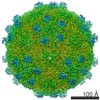 3528MC M: map data used to model this data C: citing same article ( |
|---|---|
| Similar structure data | |
| EM raw data | EMPIAR-10088 (Title: Single particle dataset of Nora virus / Data size: 3.6 TB Data #1: Unaligned multi-frame micrographs of Nora virus [micrographs - multiframe]) |
-Links
 PDBj
PDBj- Assembly
| Deposited unit | 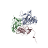
|
|---|---|
| 1 | x 60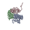
|
| Symmetry | Point symmetry: (Schoenflies symbol: I (icosahedral)) |
-Components
| #1: Protein | Mass: 45258.152 Da / Num. of mol.: 1 / Source method: isolated from a natural source / Source: (natural) Nora virus / References: UniProt: Q27YG7 |
|---|---|
| #2: Protein | Mass: 28166.934 Da / Num. of mol.: 1 / Source method: isolated from a natural source / Source: (natural) Nora virus / References: UniProt: Q27YG7 |
| #3: Protein | Mass: 29019.826 Da / Num. of mol.: 1 / Source method: isolated from a natural source / Source: (natural) Nora virus / References: UniProt: D2WFA0 |
-Experimental details
-Experiment
| Experiment | Method: ELECTRON MICROSCOPY |
|---|---|
| EM experiment | Aggregation state: PARTICLE / 3D reconstruction method: single particle reconstruction |
- Sample preparation
| Component | Name: Nora virus / Type: VIRUS Details: Drosophila Nora virus strain Umea 2007 was derived from an infectious cDNA clone (accession GQ257737). Entity ID: all / Source: RECOMBINANT |
|---|---|
| Source (natural) | Organism: Nora virus |
| Source (recombinant) | Organism:  |
| Details of virus | Empty: NO / Enveloped: NO / Isolate: SPECIES / Type: VIRION |
| Natural host | Organism: Drosophila melanogaster |
| Virus shell | Triangulation number (T number): 1 |
| Buffer solution | pH: 7.4 / Details: 10mM Tris-HCl pH 7.4. |
| Specimen | Embedding applied: NO / Shadowing applied: NO / Staining applied: NO / Vitrification applied: YES / Details: This sample was monodisperse. |
| Specimen support | Grid material: COPPER / Grid mesh size: 300 divisions/in. / Grid type: Quantifoil R2/2 |
| Vitrification | Instrument: LEICA EM GP / Cryogen name: ETHANE / Humidity: 80 % / Chamber temperature: 295 K |
- Electron microscopy imaging
| Experimental equipment |  Model: Titan Krios / Image courtesy: FEI Company |
|---|---|
| Microscopy | Model: FEI TITAN KRIOS |
| Electron gun | Electron source:  FIELD EMISSION GUN / Accelerating voltage: 300 kV / Illumination mode: FLOOD BEAM FIELD EMISSION GUN / Accelerating voltage: 300 kV / Illumination mode: FLOOD BEAM |
| Electron lens | Mode: BRIGHT FIELD / Calibrated magnification: 47170 X / Nominal defocus max: 1500 nm / Nominal defocus min: 500 nm / Cs: 2.7 mm |
| Specimen holder | Cryogen: NITROGEN / Specimen holder model: FEI TITAN KRIOS AUTOGRID HOLDER |
| Image recording | Average exposure time: 8 sec. / Electron dose: 40 e/Å2 / Detector mode: INTEGRATING / Film or detector model: GATAN K2 QUANTUM (4k x 4k) / Num. of grids imaged: 1 / Num. of real images: 3516 |
| EM imaging optics | Energyfilter name: GIF Quantum |
| Image scans | Movie frames/image: 20 |
- Processing
| EM software |
| ||||||||||||||||||||||||||||||||||||||||||||
|---|---|---|---|---|---|---|---|---|---|---|---|---|---|---|---|---|---|---|---|---|---|---|---|---|---|---|---|---|---|---|---|---|---|---|---|---|---|---|---|---|---|---|---|---|---|
| CTF correction | Type: PHASE FLIPPING AND AMPLITUDE CORRECTION | ||||||||||||||||||||||||||||||||||||||||||||
| Particle selection | Num. of particles selected: 30313 | ||||||||||||||||||||||||||||||||||||||||||||
| Symmetry | Point symmetry: I (icosahedral) | ||||||||||||||||||||||||||||||||||||||||||||
| 3D reconstruction | Resolution: 2.7 Å / Resolution method: FSC 0.143 CUT-OFF / Num. of particles: 16131 / Algorithm: BACK PROJECTION / Num. of class averages: 1 / Symmetry type: POINT | ||||||||||||||||||||||||||||||||||||||||||||
| Atomic model building | Protocol: AB INITIO MODEL / Space: REAL / Target criteria: Cross-correlation coefficient |
