 Movie
Movie Controller


+ Open data

- Basic information
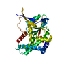
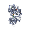
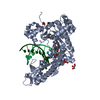
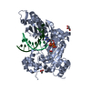
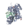
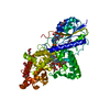
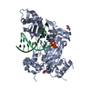
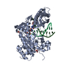
| Entry | Database: PDB / ID: 5mlq | ||||||
|---|---|---|---|---|---|---|---|
| Title | Structure of CDPS from Nocardia brasiliensis | ||||||
 Components Components | CDPS | ||||||
 Keywords Keywords | LIGASE / Cyclodipeptide Synthase | ||||||
| Function / homology | Cyclodipeptide synthase / Cyclodipeptide synthase / Cyclodipeptide synthase superfamily / aminoacyltransferase activity / CITRIC ACID / Cyclodipeptide synthase Function and homology information Function and homology information | ||||||
| Biological species |  Nocardia brasiliensis ATCC 700358 (bacteria) Nocardia brasiliensis ATCC 700358 (bacteria) | ||||||
| Method |  X-RAY DIFFRACTION / SYNCHROTRON / SAD / Resolution: 3.18 Å X-RAY DIFFRACTION / SYNCHROTRON / SAD / Resolution: 3.18 Å | ||||||
 Authors Authors | Bourgeois, G. / Seguin, J. / Moutiez, M. / Babin, M. / Belin, P. / Mechulam, Y. / Gondry, M. / Schmitt, E. | ||||||
| Funding support |  France, 1items France, 1items
| ||||||
 Citation Citation | Journal: J.Struct.Biol. / Year: 2018 Title: Structural basis for partition of the cyclodipeptide synthases into two subfamilies. Authors: Bourgeois, G. / Seguin, J. / Babin, M. / Belin, P. / Moutiez, M. / Mechulam, Y. / Gondry, M. / Schmitt, E. | ||||||
| History |
|
- Structure visualization
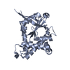
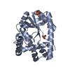
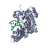
| Structure viewer | Molecule: MolmilJmol/JSmol |
|---|
- Downloads & links
-Download
| PDBx/mmCIF format | 5mlq.cif.gz | 195.5 KB | Display | PDBx/mmCIF format |
|---|---|---|---|---|
| PDB format | pdb5mlq.ent.gz | 158.7 KB | Display | PDB format |
| PDBx/mmJSON format | 5mlq.json.gz | Tree view | PDBx/mmJSON format | |
| Others |  Other downloads Other downloads |
-Validation report
| Summary document | 5mlq_validation.pdf.gz | 457.6 KB | Display | wwPDB validaton report |
|---|---|---|---|---|
| Full document | 5mlq_full_validation.pdf.gz | 463.5 KB | Display | |
| Data in XML | 5mlq_validation.xml.gz | 18.6 KB | Display | |
| Data in CIF | 5mlq_validation.cif.gz | 24.1 KB | Display | |
| Arichive directory | https://data.pdbj.org/pub/pdb/validation_reports/ml/5mlqftp://data.pdbj.org/pub/pdb/validation_reports/ml/5mlq | HTTPS FTP |
-Related structure data


-Links
 PDBj
PDBj- Assembly
| Deposited unit | 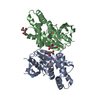
| ||||||||
|---|---|---|---|---|---|---|---|---|---|
| 1 |
| ||||||||
| Unit cell |
|
-Components
| #1: Protein | Mass: 28555.605 Da / Num. of mol.: 2 Source method: isolated from a genetically manipulated source Source: (gene. exp.) Nocardia brasiliensis ATCC 700358 (bacteria)Gene: O3I_025450 / Production host: #2: Chemical |   Mass: 192.124 Da / Num. of mol.: 3 / Source method: obtained synthetically / Formula: C6H8O7 Mass: 192.124 Da / Num. of mol.: 3 / Source method: obtained synthetically / Formula: C6H8O7Has protein modification | Y | |
|---|
-Experimental details
-Experiment
| Experiment | Method: X-RAY DIFFRACTION / Number of used crystals: 1 |
|---|
- Sample preparation
| Crystal | Density Matthews: 4.74 Å3/Da / Density % sol: 74.05 % |
|---|---|
| Crystal grow | Temperature: 278 K / Method: vapor diffusion, hanging drop / pH: 7 / Details: PEG3350 22% Tri-ammonium citrate 0.2M pH = 7 |
-Data collection
| Diffraction | Mean temperature: 100 K |
|---|---|
| Diffraction source | Source: SYNCHROTRON / Site: SOLEIL / Beamline: PROXIMA 1 / Wavelength: 0.9792 Å |
| Detector | Type: DECTRIS PILATUS3 6M / Detector: PIXEL / Date: Mar 31, 2015 |
| Radiation | Monochromator: crystal / Protocol: SINGLE WAVELENGTH / Monochromatic (M) / Laue (L): M / Scattering type: x-ray |
| Radiation wavelength | Wavelength: 0.9792 Å / Relative weight: 1 |
| Reflection | Resolution: 3.18→46.15 Å / Num. obs: 18802 / % possible obs: 99.5 % / Redundancy: 12.9 % / Biso Wilson estimate: 128.88 Å2 / CC1/2: 0.999 / Rmerge(I) obs: 0.094 / Rsym value: 0.094 / Net I/σ(I): 19.04 |
| Reflection shell | Resolution: 3.18→3.37 Å / Redundancy: 12.8 % / Rmerge(I) obs: 1.25 / Mean I/σ(I) obs: 2.05 / CC1/2: 0.846 / Rsym value: 1.25 / % possible all: 97.6 |
- Processing
| Software |
| ||||||||||||||||||||||||||||||||||||||||||||||||||||||||||||||||||||||||||||||||||||||||||||||||||||||||||||||||||
|---|---|---|---|---|---|---|---|---|---|---|---|---|---|---|---|---|---|---|---|---|---|---|---|---|---|---|---|---|---|---|---|---|---|---|---|---|---|---|---|---|---|---|---|---|---|---|---|---|---|---|---|---|---|---|---|---|---|---|---|---|---|---|---|---|---|---|---|---|---|---|---|---|---|---|---|---|---|---|---|---|---|---|---|---|---|---|---|---|---|---|---|---|---|---|---|---|---|---|---|---|---|---|---|---|---|---|---|---|---|---|---|---|---|---|---|
| Refinement | Method to determine structure: SAD / Resolution: 3.18→46.15 Å / Cor.coef. Fo:Fc: 0.953 / Cor.coef. Fo:Fc free: 0.946 / SU R Cruickshank DPI: 0.508 / Cross valid method: THROUGHOUT / σ(F): 0 / SU R Blow DPI: 0.501 / SU Rfree Blow DPI: 0.268 / SU Rfree Cruickshank DPI: 0.273
| ||||||||||||||||||||||||||||||||||||||||||||||||||||||||||||||||||||||||||||||||||||||||||||||||||||||||||||||||||
| Displacement parameters | Biso mean: 121.24 Å2
| ||||||||||||||||||||||||||||||||||||||||||||||||||||||||||||||||||||||||||||||||||||||||||||||||||||||||||||||||||
| Refine analyze | Luzzati coordinate error obs: 0.36 Å | ||||||||||||||||||||||||||||||||||||||||||||||||||||||||||||||||||||||||||||||||||||||||||||||||||||||||||||||||||
| Refinement step | Cycle: 1 / Resolution: 3.18→46.15 Å
| ||||||||||||||||||||||||||||||||||||||||||||||||||||||||||||||||||||||||||||||||||||||||||||||||||||||||||||||||||
| Refine LS restraints |
| ||||||||||||||||||||||||||||||||||||||||||||||||||||||||||||||||||||||||||||||||||||||||||||||||||||||||||||||||||
| LS refinement shell | Resolution: 3.18→3.37 Å / Rfactor Rfree error: 0 / Total num. of bins used: 9
| ||||||||||||||||||||||||||||||||||||||||||||||||||||||||||||||||||||||||||||||||||||||||||||||||||||||||||||||||||
| Refinement TLS params. | Method: refined / Refine-ID: X-RAY DIFFRACTION
| ||||||||||||||||||||||||||||||||||||||||||||||||||||||||||||||||||||||||||||||||||||||||||||||||||||||||||||||||||
| Refinement TLS group |
|
