 Movie
Movie Controller

[English] 日本語
 Yorodumi
- PDB-5mk7: Crystal structure of the receptor-binding domain of botulinum neu... -
+ Open data

- Basic information
| Entry | Database: PDB / ID: 5mk7 | ||||||
|---|---|---|---|---|---|---|---|
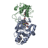
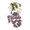
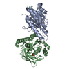
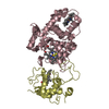
| Title | Crystal structure of the receptor-binding domain of botulinum neurotoxin A1 (crystal form 2) | ||||||
 Components Components | Botulinum neurotoxin type A | ||||||
 Keywords Keywords | TOXIN / bacterial / Toxin receptor binding domain / jelly roll fold / beta trefoil fold | ||||||
| Function / homology |  Function and homology information Function and homology informationhost cell junction / negative regulation of neurotransmitter secretion / bontoxilysin / host cell presynaptic membrane / host cell cytoplasmic vesicle / host cell cytosol / protein transmembrane transporter activity / membrane => GO:0016020 / metalloendopeptidase activity / toxin activity ...host cell junction / negative regulation of neurotransmitter secretion / bontoxilysin / host cell presynaptic membrane / host cell cytoplasmic vesicle / host cell cytosol / protein transmembrane transporter activity / membrane => GO:0016020 / metalloendopeptidase activity / toxin activity / host cell plasma membrane / proteolysis / extracellular region / zinc ion binding / membrane Similarity search - Function | ||||||
| Biological species |   Clostridium botulinum (bacteria) Clostridium botulinum (bacteria) | ||||||
| Method |  X-RAY DIFFRACTION / SYNCHROTRON / MOLECULAR REPLACEMENT / Resolution: 1.8 Å X-RAY DIFFRACTION / SYNCHROTRON / MOLECULAR REPLACEMENT / Resolution: 1.8 Å | ||||||
 Authors Authors | Davies, J.R. / Acharya, K.R. | ||||||
 Citation Citation | Journal: PeerJ / Year: 2018 Title: High resolution crystal structures of the receptor-binding domain ofClostridium botulinumneurotoxin serotypes A and FA. Authors: Davies, J.R. / Hackett, G.S. / Liu, S.M. / Acharya, K.R. | ||||||
| History |
|
- Structure visualization
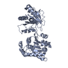
| Structure viewer | Molecule: MolmilJmol/JSmol |
|---|
- Downloads & links
-Download
| PDBx/mmCIF format | 5mk7.cif.gz | 104.3 KB | Display | PDBx/mmCIF format |
|---|---|---|---|---|
| PDB format | pdb5mk7.ent.gz | 75.6 KB | Display | PDB format |
| PDBx/mmJSON format | 5mk7.json.gz | Tree view | PDBx/mmJSON format | |
| Others |  Other downloads Other downloads |
-Validation report
| Summary document | 5mk7_validation.pdf.gz | 422.4 KB | Display | wwPDB validaton report |
|---|---|---|---|---|
| Full document | 5mk7_full_validation.pdf.gz | 425.7 KB | Display | |
| Data in XML | 5mk7_validation.xml.gz | 18.7 KB | Display | |
| Data in CIF | 5mk7_validation.cif.gz | 28 KB | Display | |
| Arichive directory | https://data.pdbj.org/pub/pdb/validation_reports/mk/5mk7ftp://data.pdbj.org/pub/pdb/validation_reports/mk/5mk7 | HTTPS FTP |
-Related structure data
| Related structure data |  5mk6C  5mk8C 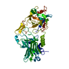 2vuaS S: Starting model for refinement C: citing same article ( |
|---|---|
| Similar structure data |


-Links
 PDBj
PDBj- Assembly
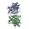
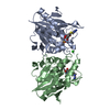
| Deposited unit | 
| ||||||||
|---|---|---|---|---|---|---|---|---|---|
| 1 |
| ||||||||
| Unit cell |
|
-Components
| #1: Protein | Mass: 50711.406 Da / Num. of mol.: 1 Source method: isolated from a genetically manipulated source Source: (gene. exp.) Clostridium botulinum (bacteria) / Gene: botA, atx, bna / Production host: References: UniProt: P10845, UniProt: P0DPI1*PLUS, bontoxilysin |
|---|---|
| #2: Water | ChemComp-HOH /  Mass: 18.015 Da / Num. of mol.: 322 / Source method: isolated from a natural source / Formula: H2O Mass: 18.015 Da / Num. of mol.: 322 / Source method: isolated from a natural source / Formula: H2O |
| Has protein modification | Y |
-Experimental details
-Experiment
| Experiment | Method: X-RAY DIFFRACTION / Number of used crystals: 1 |
|---|
- Sample preparation
| Crystal | Density Matthews: 1.97 Å3/Da / Density % sol: 37.42 % |
|---|---|
| Crystal grow | Temperature: 289.15 K / Method: vapor diffusion, sitting drop / pH: 4 / Details: 0.1 M MIB, 25% w/v PEG 1500 |
-Data collection
| Diffraction | Mean temperature: 100 K |
|---|---|
| Diffraction source | Source: SYNCHROTRON / Site: Diamond  / Beamline: I02 / Wavelength: 0.97949 Å / Beamline: I02 / Wavelength: 0.97949 Å |
| Detector | Type: DECTRIS PILATUS 6M-F / Detector: PIXEL / Date: Jun 25, 2016 |
| Radiation | Protocol: SINGLE WAVELENGTH / Monochromatic (M) / Laue (L): M / Scattering type: x-ray |
| Radiation wavelength | Wavelength: 0.97949 Å / Relative weight: 1 |
| Reflection | Resolution: 1.8→60.5 Å / Num. obs: 36131 / % possible obs: 98.6 % / Redundancy: 20.4 % / CC1/2: 0.962 / Net I/σ(I): 14.4 |
| Reflection shell | Resolution: 1.8→1.84 Å / Redundancy: 11.7 % / Mean I/σ(I) obs: 3.7 / CC1/2: 0.954 / % possible all: 89.1 |
- Processing
| Software |
| ||||||||||||||||||||||||||||||||||||||||||||||||||||||||||||||||||||||||||||||||||||||||||||||||||
|---|---|---|---|---|---|---|---|---|---|---|---|---|---|---|---|---|---|---|---|---|---|---|---|---|---|---|---|---|---|---|---|---|---|---|---|---|---|---|---|---|---|---|---|---|---|---|---|---|---|---|---|---|---|---|---|---|---|---|---|---|---|---|---|---|---|---|---|---|---|---|---|---|---|---|---|---|---|---|---|---|---|---|---|---|---|---|---|---|---|---|---|---|---|---|---|---|---|---|---|
| Refinement | Method to determine structure: MOLECULAR REPLACEMENT Starting model: 2VUA Resolution: 1.8→60.242 Å / SU ML: 0.17 / Cross valid method: FREE R-VALUE / σ(F): 1.34 / Phase error: 24.28
| ||||||||||||||||||||||||||||||||||||||||||||||||||||||||||||||||||||||||||||||||||||||||||||||||||
| Solvent computation | Shrinkage radii: 0.9 Å / VDW probe radii: 1.11 Å | ||||||||||||||||||||||||||||||||||||||||||||||||||||||||||||||||||||||||||||||||||||||||||||||||||
| Refinement step | Cycle: LAST / Resolution: 1.8→60.242 Å
| ||||||||||||||||||||||||||||||||||||||||||||||||||||||||||||||||||||||||||||||||||||||||||||||||||
| Refine LS restraints |
| ||||||||||||||||||||||||||||||||||||||||||||||||||||||||||||||||||||||||||||||||||||||||||||||||||
| LS refinement shell |
|
