 Movie
Movie Controller

[English] 日本語
 Yorodumi
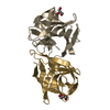
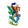
- PDB-5mh1: Crystal structure of a DM9 domain containing protein from Crassos... -
+ Open data

- Basic information
| Entry | Database: PDB / ID: 5mh1 | ||||||
|---|---|---|---|---|---|---|---|
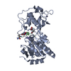
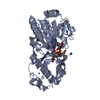
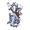
| Title | Crystal structure of a DM9 domain containing protein from Crassostrea gigas | ||||||
 Components Components | Natterin-3 | ||||||
 Keywords Keywords | SUGAR BINDING PROTEIN / beta fold | ||||||
| Function / homology | DM9 repeat / DM9 repeat / Repeats found in Drosophila proteins. / metal ion binding / beta-D-mannopyranose / Natterin-3 Function and homology information Function and homology information | ||||||
| Biological species |  Crassostrea gigas (Pacific oyster) Crassostrea gigas (Pacific oyster) | ||||||
| Method |  X-RAY DIFFRACTION / SYNCHROTRON / MOLECULAR REPLACEMENT / Resolution: 1.1 Å X-RAY DIFFRACTION / SYNCHROTRON / MOLECULAR REPLACEMENT / Resolution: 1.1 Å | ||||||
 Authors Authors | Weinert, T. / Warkentin, E. / Pang, G. | ||||||
 Citation Citation | Journal: Front Immunol / Year: 2017 Title: DM9 Domain Containing Protein Functions As a Pattern Recognition Receptor with Broad Microbial Recognition Spectrum. Authors: Jiang, S. / Wang, L. / Huang, M. / Jia, Z. / Weinert, T. / Warkentin, E. / Liu, C. / Song, X. / Zhang, H. / Witt, J. / Qiu, L. / Peng, G. / Song, L. #1: Journal: To Be PublishedTitle: A DM9 domain containing protein functions as a pattern recognition receptor with novel structural basis in Crassostrea gigas Authors: Jiang, S. / Huang, M. | ||||||
| History |
|
- Structure visualization
| Structure viewer | Molecule: MolmilJmol/JSmol |
|---|
- Downloads & links
-Download
| PDBx/mmCIF format | 5mh1.cif.gz | 105 KB | Display | PDBx/mmCIF format |
|---|---|---|---|---|
| PDB format | pdb5mh1.ent.gz | 81.2 KB | Display | PDB format |
| PDBx/mmJSON format | 5mh1.json.gz | Tree view | PDBx/mmJSON format | |
| Others |  Other downloads Other downloads |
-Validation report
| Arichive directory | https://data.pdbj.org/pub/pdb/validation_reports/mh/5mh1ftp://data.pdbj.org/pub/pdb/validation_reports/mh/5mh1 | HTTPS FTP |
|---|
-Related structure data
| Related structure data | 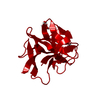 5mh0SC 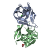 5mh2C  5mh3C S: Starting model for refinement C: citing same article ( |
|---|---|
| Similar structure data |


-Links
 PDBj
PDBj- Assembly
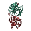
| Deposited unit | 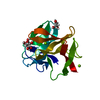
| |||||||||
|---|---|---|---|---|---|---|---|---|---|---|
| 1 | 
| |||||||||
| Unit cell |
| |||||||||
| Components on special symmetry positions |
|
-Components
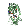
| #1: Protein | Mass: 15536.697 Da / Num. of mol.: 1 Source method: isolated from a genetically manipulated source Source: (gene. exp.) Crassostrea gigas (Pacific oyster) / Gene: CGI_10011001, CGI_10018577 / Production host:  | ||
|---|---|---|---|
| #2: Chemical | ChemComp-GOL /   Mass: 92.094 Da / Num. of mol.: 1 / Source method: obtained synthetically / Formula: C3H8O3 Mass: 92.094 Da / Num. of mol.: 1 / Source method: obtained synthetically / Formula: C3H8O3 | ||
| #3: Sugar | ChemComp-BMA /   Type: D-saccharide, beta linking / Mass: 180.156 Da / Num. of mol.: 1 Type: D-saccharide, beta linking / Mass: 180.156 Da / Num. of mol.: 1Source method: isolated from a genetically manipulated source Formula: C6H12O6 | ||
| #4: Chemical |   Mass: 24.305 Da / Num. of mol.: 2 / Source method: obtained synthetically / Formula: Mg Mass: 24.305 Da / Num. of mol.: 2 / Source method: obtained synthetically / Formula: Mg#5: Water | ChemComp-HOH / |  Mass: 18.015 Da / Num. of mol.: 234 / Source method: isolated from a natural source / Formula: H2O Mass: 18.015 Da / Num. of mol.: 234 / Source method: isolated from a natural source / Formula: H2O |
-Experimental details
-Experiment
| Experiment | Method: X-RAY DIFFRACTION / Number of used crystals: 1 |
|---|
- Sample preparation
| Crystal | Density Matthews: 2.31 Å3/Da / Density % sol: 46.85 % |
|---|---|
| Crystal grow | Temperature: 291 K / Method: vapor diffusion, sitting drop / pH: 6 / Details: 0.1M MES, pH 6.0, 20% PEG 6000, 10 mM mannose |
-Data collection
| Diffraction | Mean temperature: 100 K |
|---|---|
| Diffraction source | Source: SYNCHROTRON / Site: SRS  / Beamline: PX10.1 / Wavelength: 0.999999 Å / Beamline: PX10.1 / Wavelength: 0.999999 Å |
| Detector | Type: DECTRIS PILATUS3 S 6M / Detector: PIXEL / Date: Jul 21, 2014 |
| Radiation | Protocol: SINGLE WAVELENGTH / Monochromatic (M) / Laue (L): M / Scattering type: x-ray |
| Radiation wavelength | Wavelength: 0.999999 Å / Relative weight: 1 |
| Reflection | Resolution: 1.1→10 Å / Num. all: 985047 / Num. obs: 58075 / % possible obs: 99.9 % / Redundancy: 17 % / CC1/2: 0.99 / Rrim(I) all: 0.072 / Net I/σ(I): 21.7 |
| Reflection shell | Resolution: 1.1→1.2 Å / Redundancy: 14.5 % / Mean I/σ(I) obs: 1.4 / Num. measured obs: 184866 / Num. unique all: 12761 / CC1/2: 0.806 / Rrim(I) all: 2 / % possible all: 95.9 |
- Processing
| Software |
| ||||||||||||||||||||||||
|---|---|---|---|---|---|---|---|---|---|---|---|---|---|---|---|---|---|---|---|---|---|---|---|---|---|
| Refinement | Method to determine structure: MOLECULAR REPLACEMENT Starting model: 5MH0 Resolution: 1.1→5 Å / SU ML: 0.04 / Cross valid method: THROUGHOUT / σ(F): 1.39 / Phase error: 8.62
| ||||||||||||||||||||||||
| Solvent computation | Shrinkage radii: 0.9 Å / VDW probe radii: 1.11 Å | ||||||||||||||||||||||||
| Refinement step | Cycle: LAST / Resolution: 1.1→5 Å
| ||||||||||||||||||||||||
| Refine LS restraints |
|
