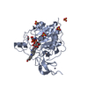
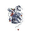
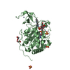
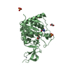
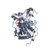
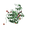
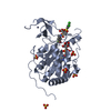
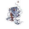
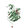
Entry Database : PDB / ID : 5mfzTitle Human Sirt6 in complex with small molecule UBCS40 NAD-dependent protein deacetylase sirtuin-6 Keywords / / / / Function / homology Function Domain/homology Component
/ / / / / / / / / / / / / / / / / / / / / / / / / / / / / / / / / / / / / / / / / / / / / / / / / / / / / / / / / / / / / / / / / / / / / / / / / / / / / / / / / / / / / / / / / / / / / / / / / / / / / / / / / / / / / / / / / / / / / Biological species Homo sapiens (human)Method / / / Resolution : 2.1 Å Authors Steegborn, C. / You, W. / Kambach, C. Funding support Organization Grant number Country Oberfrankenstiftung
Journal : Angew. Chem. Int. Ed. Engl. / Year : 2017Title : Structural Basis of Sirtuin 6 Activation by Synthetic Small Molecules.Authors : You, W. / Rotili, D. / Li, T.M. / Kambach, C. / Meleshin, M. / Schutkowski, M. / Chua, K.F. / Mai, A. / Steegborn, C. History Deposition Nov 19, 2016 Deposition site / Processing site Revision 1.0 Dec 28, 2016 Provider / Type Revision 1.1 Jan 25, 2017 Group Revision 1.2 Jan 17, 2024 Group Data collection / Database references ... Data collection / Database references / Refinement description / Structure summary Category chem_comp / chem_comp_atom ... chem_comp / chem_comp_atom / chem_comp_bond / database_2 / pdbx_initial_refinement_model Item / _database_2.pdbx_DOI / _database_2.pdbx_database_accession
Show all Show less
 Movie
Movie Controller


 Open data
 Basic information
 Components
 Keywords
 Function and homology information
 Homo sapiens (human)
 X-RAY DIFFRACTION /
  Authors
 Germany, 1items
  Citation
 Structure visualization
 Downloads & links
 Other downloads
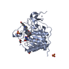


 PDBj
PDBj

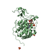
 Assembly
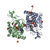
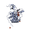

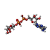


 Mass: 559.316 Da / Num. of mol.: 2 / Source method: obtained synthetically / Formula: C15H23N5O14P2
 Mass: 65.409 Da / Num. of mol.: 2 / Source method: obtained synthetically / Formula: Zn
 Mass: 96.063 Da / Num. of mol.: 8 / Source method: obtained synthetically / Formula: SO4
 Mass: 310.349 Da / Num. of mol.: 1 / Source method: obtained synthetically / Formula: C21H14N2O
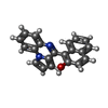
 Mass: 62.068 Da / Num. of mol.: 1 / Source method: obtained synthetically / Formula: C2H6O2
 Sample preparation
 Processing