 Movie
Movie Controller

[English] 日本語
 Yorodumi
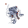
- PDB-6qch: Human Sirt6 in complex with ADP-ribose and the activator cyanidin -
+ Open data

- Basic information
| Entry | Database: PDB / ID: 6qch | ||||||
|---|---|---|---|---|---|---|---|
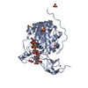
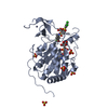
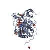
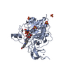
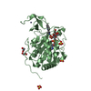
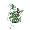
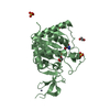
| Title | Human Sirt6 in complex with ADP-ribose and the activator cyanidin | ||||||
 Components Components | NAD-dependent protein deacetylase sirtuin-6 | ||||||
 Keywords Keywords | HYDROLASE / Deacylase / Activator / Quercetin derivative / Allosteric | ||||||
| Function / homology |  Function and homology information Function and homology informationhistone H3K56 deacetylase activity, NAD-dependent / ketone biosynthetic process / histone H3K18 deacetylase activity, NAD-dependent / histone H3K9 deacetylase activity, hydrolytic mechanism / histone H3K9 deacetylase activity, NAD-dependent / protein delipidation / NAD+-protein-lysine ADP-ribosyltransferase activity / chromosome, subtelomeric region / regulation of lipid catabolic process / positive regulation of protein localization to chromatin ...histone H3K56 deacetylase activity, NAD-dependent / ketone biosynthetic process / histone H3K18 deacetylase activity, NAD-dependent / histone H3K9 deacetylase activity, hydrolytic mechanism / histone H3K9 deacetylase activity, NAD-dependent / protein delipidation / NAD+-protein-lysine ADP-ribosyltransferase activity / chromosome, subtelomeric region / regulation of lipid catabolic process / positive regulation of protein localization to chromatin / NAD+-protein-arginine ADP-ribosyltransferase activity / pericentric heterochromatin formation / NAD-dependent protein demyristoylase activity / NAD-dependent protein depalmitoylase activity / positive regulation of stem cell differentiation / DNA damage sensor activity / negative regulation of D-glucose import across plasma membrane / positive regulation of chondrocyte proliferation / cardiac muscle cell differentiation / transposable element silencing / protein acetyllysine N-acetyltransferase / NAD-dependent protein lysine deacetylase activity / protein deacetylation / histone deacetylase activity, NAD-dependent / positive regulation of blood vessel branching / protein localization to site of double-strand break / positive regulation of vascular endothelial cell proliferation / negative regulation of glycolytic process / positive regulation of stem cell proliferation / negative regulation of protein import into nucleus / TORC2 complex binding / regulation of protein secretion / regulation of double-strand break repair via homologous recombination / lncRNA binding / negative regulation of gene expression, epigenetic / NAD+-protein mono-ADP-ribosyltransferase activity / positive regulation of stem cell population maintenance / negative regulation of cellular senescence / positive regulation of double-strand break repair / negative regulation of transcription elongation by RNA polymerase II / regulation of lipid metabolic process / positive regulation of telomere maintenance / Transferases; Glycosyltransferases; Pentosyltransferases / NAD+ poly-ADP-ribosyltransferase activity / NAD+ binding / DNA repair-dependent chromatin remodeling / subtelomeric heterochromatin formation / negative regulation of gluconeogenesis / positive regulation of fat cell differentiation / response to UV / pericentric heterochromatin / nucleosome binding / regulation of protein localization to plasma membrane / site of DNA damage / nucleotidyltransferase activity / negative regulation of protein localization to chromatin / Transferases; Acyltransferases; Transferring groups other than aminoacyl groups / positive regulation of protein export from nucleus / determination of adult lifespan / circadian regulation of gene expression / chromatin DNA binding / regulation of circadian rhythm / base-excision repair / protein import into nucleus / protein destabilization / Pre-NOTCH Transcription and Translation / positive regulation of fibroblast proliferation / positive regulation of insulin secretion / transcription corepressor activity / glucose homeostasis / double-strand break repair / positive regulation of proteasomal ubiquitin-dependent protein catabolic process / positive regulation of cold-induced thermogenesis / site of double-strand break / Processing of DNA double-strand break ends / damaged DNA binding / chromatin remodeling / negative regulation of cell population proliferation / chromatin binding / chromatin / negative regulation of transcription by RNA polymerase II / endoplasmic reticulum / protein homodimerization activity / DNA binding / zinc ion binding / nucleoplasm / nucleus Similarity search - Function | ||||||
| Biological species |  Homo sapiens (human) Homo sapiens (human) | ||||||
| Method |  X-RAY DIFFRACTION / SYNCHROTRON / MOLECULAR REPLACEMENT / Resolution: 2.1 Å X-RAY DIFFRACTION / SYNCHROTRON / MOLECULAR REPLACEMENT / Resolution: 2.1 Å | ||||||
 Authors Authors | You, W. / Steegborn, C. | ||||||
| Funding support |  Germany, 1items Germany, 1items
| ||||||
 Citation Citation | Journal: Sci Rep / Year: 2019 Title: Structural basis for the activation and inhibition of Sirtuin 6 by quercetin and its derivatives. Authors: You, W. / Zheng, W. / Weiss, S. / Chua, K.F. / Steegborn, C. | ||||||
| History |
|
- Structure visualization
| Structure viewer | Molecule: MolmilJmol/JSmol |
|---|
- Downloads & links
-Download
| PDBx/mmCIF format | 6qch.cif.gz | 131.7 KB | Display | PDBx/mmCIF format |
|---|---|---|---|---|
| PDB format | pdb6qch.ent.gz | 100.8 KB | Display | PDB format |
| PDBx/mmJSON format | 6qch.json.gz | Tree view | PDBx/mmJSON format | |
| Others |  Other downloads Other downloads |
-Validation report
| Arichive directory | https://data.pdbj.org/pub/pdb/validation_reports/qc/6qchftp://data.pdbj.org/pub/pdb/validation_reports/qc/6qch | HTTPS FTP |
|---|
-Related structure data
| Related structure data |  6qcdC  6qceC 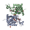 6qcjC 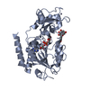 6qcnC 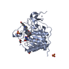 5mf6S S: Starting model for refinement C: citing same article ( |
|---|---|
| Similar structure data |


-Links
 PDBj
PDBj

- Assembly
| Deposited unit | 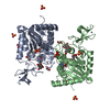
| |||||||||
|---|---|---|---|---|---|---|---|---|---|---|
| 1 | 
| |||||||||
| 2 | 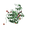
| |||||||||
| Unit cell |
| |||||||||
| Components on special symmetry positions |
|
-Components
-Protein , 1 types, 2 molecules AB
| #1: Protein | Mass: 33631.594 Da / Num. of mol.: 2 Source method: isolated from a genetically manipulated source Source: (gene. exp.) Homo sapiens (human) / Gene: SIRT6, SIR2L6 / Plasmid: pET151-D-TOPO / Production host:  References: UniProt: Q8N6T7, Hydrolases; Acting on carbon-nitrogen bonds, other than peptide bonds; In linear amides |
|---|
-Non-polymers , 6 types, 137 molecules 


| #2: Chemical |  Mass: 559.316 Da / Num. of mol.: 2 / Source method: obtained synthetically / Formula: C15H23N5O14P2 Mass: 559.316 Da / Num. of mol.: 2 / Source method: obtained synthetically / Formula: C15H23N5O14P2#3: Chemical |  Mass: 65.409 Da / Num. of mol.: 2 / Source method: obtained synthetically / Formula: Zn Mass: 65.409 Da / Num. of mol.: 2 / Source method: obtained synthetically / Formula: Zn#4: Chemical | ChemComp-EDO /  Mass: 62.068 Da / Num. of mol.: 4 / Source method: obtained synthetically / Formula: C2H6O2 Mass: 62.068 Da / Num. of mol.: 4 / Source method: obtained synthetically / Formula: C2H6O2#5: Chemical | ChemComp-SO4 /  Mass: 96.063 Da / Num. of mol.: 10 / Source method: obtained synthetically / Formula: SO4 Mass: 96.063 Da / Num. of mol.: 10 / Source method: obtained synthetically / Formula: SO4#6: Chemical |  Mass: 287.244 Da / Num. of mol.: 2 / Source method: obtained synthetically / Formula: C15H11O6 Mass: 287.244 Da / Num. of mol.: 2 / Source method: obtained synthetically / Formula: C15H11O6#7: Water | ChemComp-HOH / | Mass: 18.015 Da / Num. of mol.: 117 / Source method: isolated from a natural source / Formula: H2O |
|---|
-Experimental details
-Experiment
| Experiment | Method: X-RAY DIFFRACTION / Number of used crystals: 1 |
|---|
- Sample preparation
| Crystal | Density Matthews: 2.63 Å3/Da / Density % sol: 53.2 % |
|---|---|
| Crystal grow | Temperature: 293 K / Method: vapor diffusion, hanging drop / pH: 5.7 Details: 1.6 M (NH4)2SO4, 10% PEG 400, and Bis-Tris buffer pH 5.7 PH range: 5.7-6.2 |
-Data collection
| Diffraction | Mean temperature: 100 K / Serial crystal experiment: N | |||||||||||||||
|---|---|---|---|---|---|---|---|---|---|---|---|---|---|---|---|---|
| Diffraction source | Source: SYNCHROTRON / Site: BESSY / Beamline: 14.1 / Wavelength: 0.918 Å | |||||||||||||||
| Detector | Type: DECTRIS PILATUS 6M / Detector: PIXEL / Date: Jul 12, 2018 | |||||||||||||||
| Radiation | Protocol: SINGLE WAVELENGTH / Monochromatic (M) / Laue (L): M / Scattering type: x-ray | |||||||||||||||
| Radiation wavelength | Wavelength: 0.918 Å / Relative weight: 1 | |||||||||||||||
| Reflection twin |
| |||||||||||||||
| Reflection | Resolution: 2.1→47.94 Å / Num. obs: 39771 / % possible obs: 99.9 % / Observed criterion σ(I): 1.3 / Redundancy: 11.3 % / Biso Wilson estimate: 47.58 Å2 / CC1/2: 0.998 / Rrim(I) all: 0.16 / Net I/σ(I): 11 | |||||||||||||||
| Reflection shell | Resolution: 2.1→2.22 Å / Redundancy: 11.3 % / Mean I/σ(I) obs: 1.3 / Num. unique obs: 6379 / CC1/2: 0.634 / Rrim(I) all: 1.84 / % possible all: 99.5 |
- Processing
| Software |
| ||||||||||||||||||||||||||||||||||||||||||||||||||||||||||||||||||||||||||||||||||||||||||||||||||||||||||||||||||||||||||||||||||||||||||||||||||||||||||||||||||||||||||||||||||||||
|---|---|---|---|---|---|---|---|---|---|---|---|---|---|---|---|---|---|---|---|---|---|---|---|---|---|---|---|---|---|---|---|---|---|---|---|---|---|---|---|---|---|---|---|---|---|---|---|---|---|---|---|---|---|---|---|---|---|---|---|---|---|---|---|---|---|---|---|---|---|---|---|---|---|---|---|---|---|---|---|---|---|---|---|---|---|---|---|---|---|---|---|---|---|---|---|---|---|---|---|---|---|---|---|---|---|---|---|---|---|---|---|---|---|---|---|---|---|---|---|---|---|---|---|---|---|---|---|---|---|---|---|---|---|---|---|---|---|---|---|---|---|---|---|---|---|---|---|---|---|---|---|---|---|---|---|---|---|---|---|---|---|---|---|---|---|---|---|---|---|---|---|---|---|---|---|---|---|---|---|---|---|---|---|
| Refinement | Method to determine structure: MOLECULAR REPLACEMENT Starting model: 5MF6 Resolution: 2.1→45.69 Å / Cor.coef. Fo:Fc: 0.967 / Cor.coef. Fo:Fc free: 0.953 / Cross valid method: THROUGHOUT / ESU R: 0.036 / ESU R Free: 0.032 / Details: HYDROGENS HAVE BEEN ADDED IN THE RIDING POSITIONS
| ||||||||||||||||||||||||||||||||||||||||||||||||||||||||||||||||||||||||||||||||||||||||||||||||||||||||||||||||||||||||||||||||||||||||||||||||||||||||||||||||||||||||||||||||||||||
| Solvent computation | Ion probe radii: 0.8 Å / Shrinkage radii: 0.8 Å / VDW probe radii: 1.2 Å | ||||||||||||||||||||||||||||||||||||||||||||||||||||||||||||||||||||||||||||||||||||||||||||||||||||||||||||||||||||||||||||||||||||||||||||||||||||||||||||||||||||||||||||||||||||||
| Displacement parameters | Biso mean: 50.493 Å2
| ||||||||||||||||||||||||||||||||||||||||||||||||||||||||||||||||||||||||||||||||||||||||||||||||||||||||||||||||||||||||||||||||||||||||||||||||||||||||||||||||||||||||||||||||||||||
| Refinement step | Cycle: 1 / Resolution: 2.1→45.69 Å
| ||||||||||||||||||||||||||||||||||||||||||||||||||||||||||||||||||||||||||||||||||||||||||||||||||||||||||||||||||||||||||||||||||||||||||||||||||||||||||||||||||||||||||||||||||||||
| Refine LS restraints |
|