 Movie
Movie Controller

[English] 日本語
 Yorodumi
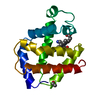
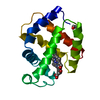
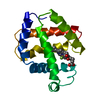
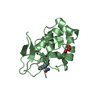
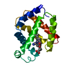
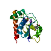
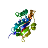
- PDB-5mba: BINDING MODE OF AZIDE TO FERRIC APLYSIA LIMACINA MYOGLOBIN. CRYST... -
+ Open data

- Basic information
| Entry | Database: PDB / ID: 5mba | |||||||||
|---|---|---|---|---|---|---|---|---|---|---|
| Title | BINDING MODE OF AZIDE TO FERRIC APLYSIA LIMACINA MYOGLOBIN. CRYSTALLOGRAPHIC ANALYSIS AT 1.9 ANGSTROMS RESOLUTION | |||||||||
 Components Components | MYOGLOBIN | |||||||||
 Keywords Keywords | OXYGEN STORAGE | |||||||||
| Function / homology |  Function and homology information Function and homology informationhemoglobin complex / oxygen carrier activity / oxygen binding / oxidoreductase activity / iron ion binding / heme binding / extracellular region Similarity search - Function | |||||||||
| Biological species |  | |||||||||
| Method |  X-RAY DIFFRACTION / Resolution: 1.9 Å X-RAY DIFFRACTION / Resolution: 1.9 Å | |||||||||
 Authors Authors | Bolognesi, M. / Onesti, S. / Gatti, G. / Coda, A. / Ascenzi, P. / Brunori, M. | |||||||||
 Citation Citation | Journal: J.Mol.Recog. / Year: 1991 Title: Binding mode of azide to ferric Aplysia limacina myoglobin. Crystallographic analysis at 1.9 A resolution. Authors: Mattevi, A. / Gatti, G. / Coda, A. / Rizzi, M. / Ascenzi, P. / Brunori, M. / Bolognesi, M. #1: Journal: Structure and Function of Invertebrate Oxygen CarriersYear: 1991 Title: Aplysia Limacina Myoglobin: Molecular Bases for Ligand Binding Authors: Bolognesi, M. / Frigerio, F. / Lionetti, C. / Rizzi, M. / Ascenzi, P. / Brunori, M. #2: Journal: J.Mol.Biol. / Year: 1989Title: Aplysia Limacina Myoglobin. Crystallographic Analysis at 1.6 Angstroms Resolution Authors: Bolognesi, M. / Onesti, S. / Gatti, G. / Coda, A. / Ascenzi, P. / Brunori, M. #3: Journal: J.Mol.Biol. / Year: 1985Title: Crystal Structure of Ferric Aplysia Limacina Myoglobin at 2.0 Angstroms Resolution Authors: Bolognesi, M. / Coda, A. / Gatti, G. / Ascenzi, P. / Brunori, M. #4: Journal: J.Mol.Biol. / Year: 1982Title: Reactivity of Ferric Aplysia and Sperm Whale Myoglobins Towards Imidazole. X-Ray and Binding Study Authors: Bolognesi, M. / Cannillo, E. / Ascenzi, P. / Giacometti, G.M. / Merli, A. / Brunori, M. #5: Journal: Acta Crystallogr.,Sect.A (Supplement) / Year: 1978Title: The Structure of Aplysia Limacina Myoglobin at 3.6 Angstroms Resolution Authors: Bolognesi, M. / Cannillo, E. / Oberti, R. / Rossi, G. / Ungaretti, L. #6: Journal: Acta Crystallogr.,Sect.B / Year: 1978Title: The Crystal Structure of met-Myoglobin from Aplysia Limacina at 5 Angstroms Resolution Authors: Ungaretti, L. / Bolognesi, M. / Cannillo, E. / Oberti, R. / Rossi, G. #7: Journal: J.Mol.Biol. / Year: 1975Title: Crystallization and Preliminary X-Ray Diffraction Studies on met-Myoglobin from Aplysia Limacina Authors: Blundell, T.L. / Brunori, M. / Curti, B. / Bolognesi, M. / Coda, A. / Fumagalli, M. / Ungaretti, L. #8: Journal: Int.J.Pept.Protein Res. / Year: 1973Title: The Amino Acid Sequence of Myoglobin from the Mollusc Aplysia Limacina Authors: Tentori, L. / Vivaldi, G. / Carta, S. / Marinucci, M. / Massa, A. / Antonini, E. / Brunori, M. | |||||||||
| History |
|
- Structure visualization
| Structure viewer | Molecule: MolmilJmol/JSmol |
|---|
- Downloads & links
-Download
| PDBx/mmCIF format | 5mba.cif.gz | 44.9 KB | Display | PDBx/mmCIF format |
|---|---|---|---|---|
| PDB format | pdb5mba.ent.gz | 31.1 KB | Display | PDB format |
| PDBx/mmJSON format | 5mba.json.gz | Tree view | PDBx/mmJSON format | |
| Others |  Other downloads Other downloads |
-Validation report
| Arichive directory | https://data.pdbj.org/pub/pdb/validation_reports/mb/5mbaftp://data.pdbj.org/pub/pdb/validation_reports/mb/5mba | HTTPS FTP |
|---|
-Related structure data
| Similar structure data |
|---|
-Links
 PDBj
PDBj


- Assembly
| Deposited unit | 
| ||||||||
|---|---|---|---|---|---|---|---|---|---|
| 1 |
| ||||||||
| Unit cell |
|
-Components
| #1: Protein | Mass: 15349.438 Da / Num. of mol.: 1 Source method: isolated from a genetically manipulated source Source: (gene. exp.) |
|---|---|
| #2: Chemical | ChemComp-AZI /   Mass: 42.020 Da / Num. of mol.: 1 / Source method: obtained synthetically / Formula: N3 Mass: 42.020 Da / Num. of mol.: 1 / Source method: obtained synthetically / Formula: N3 |
| #3: Chemical | ChemComp-HEM /   Mass: 616.487 Da / Num. of mol.: 1 / Source method: obtained synthetically / Formula: C34H32FeN4O4 Mass: 616.487 Da / Num. of mol.: 1 / Source method: obtained synthetically / Formula: C34H32FeN4O4 |
| #4: Water | ChemComp-HOH /  Mass: 18.015 Da / Num. of mol.: 126 / Source method: isolated from a natural source / Formula: H2O Mass: 18.015 Da / Num. of mol.: 126 / Source method: isolated from a natural source / Formula: H2O |
| Has protein modification | Y |
| Nonpolymer details | AZIDE ION IS BOUND TO THE HEME IRON AT THE DISTAL SITE AND TRIGGERS A LARGE CONFORMATIONAL CHANGE ...AZIDE ION IS BOUND TO THE HEME IRON AT THE DISTAL SITE AND TRIGGERS A LARGE CONFORMATI |
-Experimental details
-Experiment
| Experiment | Method: X-RAY DIFFRACTION |
|---|
- Sample preparation
| Crystal | Density Matthews: 1.98 Å3/Da / Density % sol: 37.94 % | |||||||||||||||
|---|---|---|---|---|---|---|---|---|---|---|---|---|---|---|---|---|
| Crystal grow | *PLUS pH: 7 / Method: vapor diffusion / Details: took Bolognesi et al., from original paper | |||||||||||||||
| Components of the solutions | *PLUS
|
-Data collection
| Reflection | *PLUS Highest resolution: 1.9 Å |
|---|
- Processing
| Software | Name: TNT / Classification: refinement | ||||||||||||||||||||||||||||||
|---|---|---|---|---|---|---|---|---|---|---|---|---|---|---|---|---|---|---|---|---|---|---|---|---|---|---|---|---|---|---|---|
| Refinement | Rfactor obs: 0.139 / Highest resolution: 1.9 Å | ||||||||||||||||||||||||||||||
| Refinement step | Cycle: LAST / Highest resolution: 1.9 Å
| ||||||||||||||||||||||||||||||
| Refine LS restraints |
| ||||||||||||||||||||||||||||||
| Software | *PLUS Name: TNT / Classification: refinement | ||||||||||||||||||||||||||||||
| Refinement | *PLUS Lowest resolution: 11 Å / Num. reflection obs: 7487 / Rfactor obs: 0.139 | ||||||||||||||||||||||||||||||
| Solvent computation | *PLUS | ||||||||||||||||||||||||||||||
| Displacement parameters | *PLUS | ||||||||||||||||||||||||||||||
| Refine LS restraints | *PLUS Type: t_angle_d / Dev ideal: 2.35 |
