 Movie
Movie Controller

[English] 日本語
 Yorodumi
- PDB-5m95: STAPHYLOCOCCUS CAPITIS DIVALENT METAL ION TRANSPORTER (DMT) IN CO... -
+ Open data

- Basic information
| Entry | Database: PDB / ID: 5m95 | |||||||||
|---|---|---|---|---|---|---|---|---|---|---|
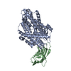
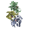
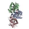
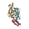
| Title | STAPHYLOCOCCUS CAPITIS DIVALENT METAL ION TRANSPORTER (DMT) IN COMPLEX WITH MANGANESE | |||||||||
 Components Components |
| |||||||||
 Keywords Keywords | TRANSPORT PROTEIN / MEMBRANE PROTEIN / TRANSPORTER / SLC11 / TRANSITION METAL IONS / NRAMP / DMT / LEUT FOLD | |||||||||
| Function / homology |  Function and homology information Function and homology informationmetal ion transmembrane transporter activity / symporter activity / membrane => GO:0016020 / metal ion binding / plasma membrane Similarity search - Function | |||||||||
| Biological species |  Staphylococcus capitis (bacteria) Staphylococcus capitis (bacteria) | |||||||||
| Method |  X-RAY DIFFRACTION / SYNCHROTRON / MOLECULAR REPLACEMENT / Resolution: 3.4 Å X-RAY DIFFRACTION / SYNCHROTRON / MOLECULAR REPLACEMENT / Resolution: 3.4 Å | |||||||||
 Authors Authors | Ehrnstorfer, I.A. / Geertsma, E.R. / Pardon, E. / Steyaert, J. / Dutzler, R. | |||||||||
 Citation Citation | Journal: Nat.Struct.Mol.Biol. / Year: 2014 Title: Crystal Structure Of A Slc11 (Nramp) Transporter Reveals The Basis For Transition-Metal Ion Transport. Authors: Ehrnstorfer, I.A. / Geertsma, E.R. / Pardon, E. / Steyaert, J. / Dutzler, R. | |||||||||
| History |
|
- Structure visualization
| Structure viewer | Molecule: MolmilJmol/JSmol |
|---|
- Downloads & links
-Download
| PDBx/mmCIF format | 5m95.cif.gz | 417.9 KB | Display | PDBx/mmCIF format |
|---|---|---|---|---|
| PDB format | pdb5m95.ent.gz | 345.9 KB | Display | PDB format |
| PDBx/mmJSON format | 5m95.json.gz | Tree view | PDBx/mmJSON format | |
| Others |  Other downloads Other downloads |
-Validation report
| Arichive directory | https://data.pdbj.org/pub/pdb/validation_reports/m9/5m95ftp://data.pdbj.org/pub/pdb/validation_reports/m9/5m95 | HTTPS FTP |
|---|
-Related structure data
| Related structure data |  5m94C  4wgv S: Starting model for refinement C: citing same article ( |
|---|---|
| Similar structure data |


-Links
 PDBj
PDBj

- Assembly
| Deposited unit | 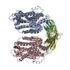
| |||||||||||||||||||||||||||||||||
|---|---|---|---|---|---|---|---|---|---|---|---|---|---|---|---|---|---|---|---|---|---|---|---|---|---|---|---|---|---|---|---|---|---|---|
| 1 | 
| |||||||||||||||||||||||||||||||||
| 2 | 
| |||||||||||||||||||||||||||||||||
| Unit cell |
| |||||||||||||||||||||||||||||||||
| Noncrystallographic symmetry (NCS) | NCS domain:
NCS domain segments:
NCS ensembles :
|
-Components
| #1: Protein | Mass: 45521.875 Da / Num. of mol.: 2 / Fragment: UNP RESIDUES 43-448 Source method: isolated from a genetically manipulated source Source: (gene. exp.) Staphylococcus capitis (bacteria)Gene: mntH, BN1317_80025, SCAPIOD100025, SCAPIOD110024, SCAPIOD130025 Production host: #2: Antibody | Mass: 13526.955 Da / Num. of mol.: 2 Source method: isolated from a genetically manipulated source Source: (gene. exp.) #3: Chemical |   Mass: 54.938 Da / Num. of mol.: 2 / Source method: obtained synthetically / Formula: Mn Mass: 54.938 Da / Num. of mol.: 2 / Source method: obtained synthetically / Formula: MnHas protein modification | Y | |
|---|
-Experimental details
-Experiment
| Experiment | Method: X-RAY DIFFRACTION / Number of used crystals: 1 |
|---|
- Sample preparation
| Crystal | Density Matthews: 4.3 Å3/Da / Density % sol: 71.36 % |
|---|---|
| Crystal grow | Temperature: 277 K / Method: vapor diffusion, sitting drop / pH: 6 / Details: 24 % PEG 400, 200 MM CACL2, 50 MM HEPES, PH 7.4 |
-Data collection
| Diffraction | Mean temperature: 100 K |
|---|---|
| Diffraction source | Source: SYNCHROTRON / Site: SLS  / Beamline: X06SA / Wavelength: 1.894 Å / Beamline: X06SA / Wavelength: 1.894 Å |
| Detector | Type: DECTRIS PILATUS 6M / Detector: PIXEL / Date: Sep 12, 2013 |
| Radiation | Protocol: SINGLE WAVELENGTH / Monochromatic (M) / Laue (L): M / Scattering type: x-ray |
| Radiation wavelength | Wavelength: 1.894 Å / Relative weight: 1 |
| Reflection | Resolution: 3.4→50 Å / Num. obs: 27350 / % possible obs: 99.9 % / Redundancy: 9.6 % / Biso Wilson estimate: 137.9 Å2 / Rmerge(I) obs: 0.081 / Net I/σ(I): 19 |
| Reflection shell | Resolution: 3.4→3.6 Å / Redundancy: 9.4 % / Rmerge(I) obs: 1.27 / Mean I/σ(I) obs: 2.4 / % possible all: 100 |
- Processing
| Software |
| |||||||||||||||||||||||||||||||||||||||||||||||||||||||||||||||||||||||||||||
|---|---|---|---|---|---|---|---|---|---|---|---|---|---|---|---|---|---|---|---|---|---|---|---|---|---|---|---|---|---|---|---|---|---|---|---|---|---|---|---|---|---|---|---|---|---|---|---|---|---|---|---|---|---|---|---|---|---|---|---|---|---|---|---|---|---|---|---|---|---|---|---|---|---|---|---|---|---|---|
| Refinement | Method to determine structure: MOLECULAR REPLACEMENT Starting model: 4WGV 4wgv Resolution: 3.4→19.9 Å / SU ML: 0.42 / Cross valid method: FREE R-VALUE / σ(F): 1.9 / Phase error: 28.37
| |||||||||||||||||||||||||||||||||||||||||||||||||||||||||||||||||||||||||||||
| Solvent computation | Shrinkage radii: 0.9 Å / VDW probe radii: 1.11 Å | |||||||||||||||||||||||||||||||||||||||||||||||||||||||||||||||||||||||||||||
| Displacement parameters | Biso mean: 152.5 Å2 | |||||||||||||||||||||||||||||||||||||||||||||||||||||||||||||||||||||||||||||
| Refinement step | Cycle: LAST / Resolution: 3.4→19.9 Å
| |||||||||||||||||||||||||||||||||||||||||||||||||||||||||||||||||||||||||||||
| Refine LS restraints |
| |||||||||||||||||||||||||||||||||||||||||||||||||||||||||||||||||||||||||||||
| Refine LS restraints NCS |
| |||||||||||||||||||||||||||||||||||||||||||||||||||||||||||||||||||||||||||||
| LS refinement shell |
| |||||||||||||||||||||||||||||||||||||||||||||||||||||||||||||||||||||||||||||
| Refinement TLS params. | Method: refined / Origin x: -2.1586 Å / Origin y: 114.0283 Å / Origin z: -13.1885 Å
| |||||||||||||||||||||||||||||||||||||||||||||||||||||||||||||||||||||||||||||
| Refinement TLS group | Selection details: ALL |
