[English] 日本語
Yorodumi
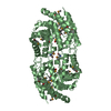
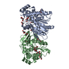
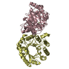
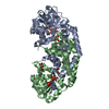
- PDB-5m8e: Crystal structure of a GH43 arabonofuranosidase from Weissella sp... -

+
Open data


ID or keywords:

Loading...

-
Basic information

Entry
Database: PDB / ID: 5m8e
Title
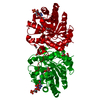
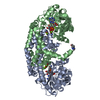
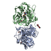
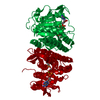
Crystal structure of a GH43 arabonofuranosidase from Weissella sp. strain 142
ComponentsAlpha-N-arabinofuranosidase
KeywordsHYDROLASE / arabinofuranosidase / 5-bladed beta-propeller fold / family 43
Function / homology
Function and homology information


non-reducing end alpha-L-arabinofuranosidase / alpha-L-arabinofuranosidase activity / carbohydrate metabolic process / metal ion binding
Similarity search - Function
Alpha-L-arabinofuranosidase / Glycoside hydrolase, family 43 / Glycosyl hydrolases family 43 / Glycosyl hydrolase domain; family 43 / 5 Propeller / Tachylectin-2; Chain A / Glycosyl hydrolase, five-bladed beta-propellor domain superfamily / Mainly Beta
Similarity search - Domain/homology
IMIDAZOLE / Alpha-N-arabinofuranosidase
Similarity search - Component
Biological speciesWeissella cibaria (bacteria)
MethodX-RAY DIFFRACTION / SYNCHROTRON / MOLECULAR REPLACEMENT / Resolution: 2 Å
AuthorsLinares-Pasten, J. / Logan, D.T. / Nordberg Karlsson, E.
Funding support Sweden, 1items
OrganizationGrant numberCountry
Swedish Research Council Sweden
CitationJournal: FEBS J. / Year: 2017
Title: Three-dimensional structures and functional studies of two GH43 arabinofuranosidases from Weissella sp. strain 142 and Lactobacillus brevis.
Authors: Linares-Pasten, J.A. / Falck, P. / Albasri, K. / Kjellstrom, S. / Adlercreutz, P. / Logan, D.T. / Karlsson, E.N.
History
DepositionOct 28, 2016Deposition site: PDBE / Processing site: PDBE
Revision 1.0May 10, 2017Provider: repository / Type: Initial release
Revision 1.1May 17, 2017Group: Database references
Revision 1.2Jul 12, 2017Group: Data collection / Database references / Category: citation / diffrn_source
Item: _citation.journal_volume / _citation.page_first ..._citation.journal_volume / _citation.page_first / _citation.page_last / _diffrn_source.pdbx_synchrotron_site
Revision 1.3Sep 13, 2017Group: Author supporting evidence / Category: pdbx_audit_support / Item: _pdbx_audit_support.funding_organization
Revision 1.4Jan 17, 2024Group: Data collection / Database references ...Data collection / Database references / Derived calculations / Refinement description
Category: chem_comp_atom / chem_comp_bond ...chem_comp_atom / chem_comp_bond / database_2 / pdbx_initial_refinement_model / pdbx_struct_conn_angle / struct_conn
Item: _database_2.pdbx_DOI / _database_2.pdbx_database_accession ..._database_2.pdbx_DOI / _database_2.pdbx_database_accession / _pdbx_struct_conn_angle.ptnr1_auth_seq_id / _pdbx_struct_conn_angle.ptnr3_auth_seq_id / _pdbx_struct_conn_angle.value / _struct_conn.pdbx_dist_value / _struct_conn.ptnr1_auth_asym_id / _struct_conn.ptnr1_auth_comp_id / _struct_conn.ptnr1_auth_seq_id / _struct_conn.ptnr1_label_asym_id / _struct_conn.ptnr1_label_atom_id / _struct_conn.ptnr1_label_comp_id / _struct_conn.ptnr1_label_seq_id / _struct_conn.ptnr2_auth_asym_id / _struct_conn.ptnr2_auth_comp_id / _struct_conn.ptnr2_auth_seq_id / _struct_conn.ptnr2_label_asym_id / _struct_conn.ptnr2_label_atom_id / _struct_conn.ptnr2_label_comp_id

-
Structure visualization

Structure viewerMolecule:
MolmilJmol/JSmol

Downloads & links

-
Assembly

Deposited unit
A: Alpha-N-arabinofuranosidase
B: Alpha-N-arabinofuranosidase
hetero molecules


Theoretical massNumber of molelcules
Total (without water)80,1259
Polymers79,6302
Non-polymers4957
Water11,458636
1


  • Idetical with deposited unit
  • defined by author
TypeNameSymmetry operationNumber
identity operation1_555x,y,z1
Unit cell
Length a, b, c (Å)59.880, 71.930, 79.510
Angle α, β, γ (deg.)90.00, 101.93, 90.00
Int Tables number4
Space group name H-MP1211
Noncrystallographic symmetry (NCS)NCS oper:
IDCodeMatrixVector
1given(1), (1), (1)
2given(-0.99809, -0.061679, 0.003525), (-0.061707, 0.998057, -0.008584), (-0.002989, -0.008785, -0.999957)74.05005, 2.65909, 78.10181

-
Components

#1: Protein Alpha-N-arabinofuranosidase / Extracellular exo-alpha-(1->5)-L-arabinofuranosidase


Mass: 39815.164 Da / Num. of mol.: 2
Source method: isolated from a genetically manipulated source
Source: (gene. exp.) Weissella cibaria (bacteria) / Gene: AO080_02990, QX99_00224 / Production host: Escherichia coli BL21(DE3) (bacteria)
References: UniProt: A0A0D1M7L2, non-reducing end alpha-L-arabinofuranosidase
#2: Chemical ChemComp-CA / CALCIUM ION


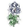
Mass: 40.078 Da / Num. of mol.: 2 / Source method: obtained synthetically / Formula: Ca
#3: Chemical ChemComp-IMD / IMIDAZOLE


Mass: 69.085 Da / Num. of mol.: 2 / Source method: obtained synthetically / Formula: C3H5N2
#4: Chemical ChemComp-GOL / GLYCEROL / GLYCERIN / PROPANE-1,2,3-TRIOL


Mass: 92.094 Da / Num. of mol.: 3 / Source method: obtained synthetically / Formula: C3H8O3
#5: Water ChemComp-HOH / water


Mass: 18.015 Da / Num. of mol.: 636 / Source method: isolated from a natural source / Formula: H2O

-
Experimental details

-
Experiment

ExperimentMethod: X-RAY DIFFRACTION / Number of used crystals: 1

-
Sample preparation

CrystalDensity Matthews: 2.11 Å3/Da / Density % sol: 41.6 %
Crystal growTemperature: 277 K / Method: vapor diffusion, sitting drop
Details: 27 to 29 % PEG 1500, 60 to 140 mM malonate-imidazole-borate (MIB) buffer, pH 4.0 to 4.5
PH range: 4.0-4.5

-
Data collection

DiffractionMean temperature: 100 K
Diffraction sourceSource: SYNCHROTRON / Site: MAX II / Beamline: I911-2 / Wavelength: 1.0384 Å
DetectorType: MAR CCD 165 mm / Detector: CCD / Date: Dec 5, 2013 / Details: focusing mirrors
RadiationMonochromator: Si(111) double crystal / Protocol: SINGLE WAVELENGTH / Monochromatic (M) / Laue (L): M / Scattering type: x-ray
Radiation wavelengthWavelength: 1.0384 Å / Relative weight: 1
ReflectionResolution: 2→29.7 Å / Num. all: 139952 / % possible obs: 98.2 % / Redundancy: 3.2 % / Biso Wilson estimate: 24.53 Å2 / Rmerge(I) obs: 0.069 / Net I/σ(I): 13.1
Reflection shellResolution: 2→2.11 Å / Redundancy: 3 % / Rmerge(I) obs: 0.395 / Mean I/σ(I) obs: 3.4 / % possible all: 92.4

-
Processing

Software
NameVersionClassification
BUSTER2.10.3refinement
XDSdata reduction
SCALAdata scaling
PHASERphasing
RefinementMethod to determine structure: MOLECULAR REPLACEMENT
Starting model: 3AKH
Resolution: 2→29.7 Å / Cor.coef. Fo:Fc: 0.957 / Cor.coef. Fo:Fc free: 0.928 / SU R Cruickshank DPI: 0.19 / Cross valid method: THROUGHOUT / σ(F): 0 / SU R Blow DPI: 0.205 / SU Rfree Blow DPI: 0.164 / SU Rfree Cruickshank DPI: 0.16
RfactorNum. reflection% reflectionSelection details
Rfree0.216 2232 5.09 %RANDOM
Rwork0.172 ---
obs0.175 43956 98 %-
Displacement parametersBiso mean: 24.58 Å2
Baniso -1Baniso -2Baniso -3
1-1.2908 Å20 Å2-1.0635 Å2
2---3.2596 Å20 Å2
3---1.9688 Å2
Refine analyzeLuzzati coordinate error obs: 0.21 Å
Refinement stepCycle: LAST / Resolution: 2→29.7 Å
ProteinNucleic acidLigandSolventTotal
Num. atoms5306 0 30 637 5973
Refine LS restraints
Refine-IDTypeDev idealNumberRestraint functionWeight
X-RAY DIFFRACTIONt_bond_d0.015547HARMONIC2
X-RAY DIFFRACTIONt_angle_deg1.037576HARMONIC2
X-RAY DIFFRACTIONt_dihedral_angle_d1824SINUSOIDAL2
X-RAY DIFFRACTIONt_incorr_chiral_ct
X-RAY DIFFRACTIONt_pseud_angle
X-RAY DIFFRACTIONt_trig_c_planes158HARMONIC2
X-RAY DIFFRACTIONt_gen_planes804HARMONIC5
X-RAY DIFFRACTIONt_it5547HARMONIC20
X-RAY DIFFRACTIONt_nbd0SEMIHARMONIC5
X-RAY DIFFRACTIONt_omega_torsion3.93
X-RAY DIFFRACTIONt_other_torsion16.85
X-RAY DIFFRACTIONt_improper_torsion
X-RAY DIFFRACTIONt_chiral_improper_torsion679SEMIHARMONIC5
X-RAY DIFFRACTIONt_sum_occupancies
X-RAY DIFFRACTIONt_utility_distance
X-RAY DIFFRACTIONt_utility_angle
X-RAY DIFFRACTIONt_utility_torsion
X-RAY DIFFRACTIONt_ideal_dist_contact6804SEMIHARMONIC4
LS refinement shellResolution: 2→2.05 Å / Total num. of bins used: 20
RfactorNum. reflection% reflection
Rfree0.276 139 5.02 %
Rwork0.1997 2629 -
all0.2035 2768 -
obs--84.18 %
Refinement TLS params.

Method: refined / Refine-ID: X-RAY DIFFRACTION

IDL112)L122)L132)L222)L232)L332)S11 (Å °)S12 (Å °)S13 (Å °)S21 (Å °)S22 (Å °)S23 (Å °)S31 (Å °)S32 (Å °)S33 (Å °)T112)T122)T132)T222)T232)T332)Origin x (Å)Origin y (Å)Origin z (Å)
10.4798-0.0863-0.20.3019-0.07571.15720.0277-0.11530.03910.0493-0.0045-0.0396-0.14640.2465-0.0232-0.0394-0.05010.0194-0.0489-0.0022-0.02839.423721.613957.2206
20.61320.2006-0.20330.5009-0.0120.93660.00540.10780.0553-0.04980.03070.0475-0.0292-0.0211-0.0361-0.03280.01610.0184-0.09140.0121-0.009233.298821.221220.5744
Refinement TLS group
IDRefine-IDRefine TLS-IDSelection details
1X-RAY DIFFRACTION1{ A|2 - 401 }
2X-RAY DIFFRACTION2{ B|2 - 401 }

+
About Yorodumi

-
News

-
Feb 9, 2022. New format data for meta-information of EMDB entries

New format data for meta-information of EMDB entries

  • Version 3 of the EMDB header file is now the official format.
  • The previous official version 1.9 will be removed from the archive.

Related info.:EMDB header

External links:wwPDB to switch to version 3 of the EMDB data model

-
Aug 12, 2020. Covid-19 info

Covid-19 info

URL: https://pdbj.org/emnavi/covid19.php

New page: Covid-19 featured information page in EM Navigator.

Related info.:Covid-19 info / Mar 5, 2020. Novel coronavirus structure data

+
Mar 5, 2020. Novel coronavirus structure data

Novel coronavirus structure data

Related info.:Yorodumi Speices / Aug 12, 2020. Covid-19 info

External links:COVID-19 featured content - PDBj / Molecule of the Month (242):Coronavirus Proteases

+
Jan 31, 2019. EMDB accession codes are about to change! (news from PDBe EMDB page)

EMDB accession codes are about to change! (news from PDBe EMDB page)

  • The allocation of 4 digits for EMDB accession codes will soon come to an end. Whilst these codes will remain in use, new EMDB accession codes will include an additional digit and will expand incrementally as the available range of codes is exhausted. The current 4-digit format prefixed with “EMD-” (i.e. EMD-XXXX) will advance to a 5-digit format (i.e. EMD-XXXXX), and so on. It is currently estimated that the 4-digit codes will be depleted around Spring 2019, at which point the 5-digit format will come into force.
  • The EM Navigator/Yorodumi systems omit the EMD- prefix.

Related info.:Q: What is EMD? / ID/Accession-code notation in Yorodumi/EM Navigator

External links:EMDB Accession Codes are Changing Soon! / Contact to PDBj

+
Jul 12, 2017. Major update of PDB

Major update of PDB

  • wwPDB released updated PDB data conforming to the new PDBx/mmCIF dictionary.
  • This is a major update changing the version number from 4 to 5, and with Remediation, in which all the entries are updated.
  • In this update, many items about electron microscopy experimental information are reorganized (e.g. em_software).
  • Now, EM Navigator and Yorodumi are based on the updated data.

External links:wwPDB Remediation / Enriched Model Files Conforming to OneDep Data Standards Now Available in the PDB FTP Archive

-
Yorodumi

Thousand views of thousand structures

  • Yorodumi is a browser for structure data from EMDB, PDB, SASBDB, etc.
  • This page is also the successor to EM Navigator detail page, and also detail information page/front-end page for Omokage search.
  • The word "yorodu" (or yorozu) is an old Japanese word meaning "ten thousand". "mi" (miru) is to see.

Related info.:EMDB / PDB / SASBDB / Comparison of 3 databanks / Yorodumi Search / Aug 31, 2016. New EM Navigator & Yorodumi / Yorodumi Papers / Jmol/JSmol / Function and homology information / Changes in new EM Navigator and Yorodumi

Read more