 Movie
Movie Controller

[English] 日本語
 Yorodumi
- PDB-5m47: Crystal structure of DapF from Corynebacterium glutamicum in comp... -
+ Open data

- Basic information
| Entry | Database: PDB / ID: 5m47 | |||||||||
|---|---|---|---|---|---|---|---|---|---|---|
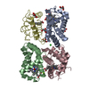
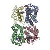
| Title | Crystal structure of DapF from Corynebacterium glutamicum in complex with D,L-diaminopimelate | |||||||||
 Components Components | Diaminopimelate epimerase | |||||||||
 Keywords Keywords | ISOMERASE | |||||||||
| Function / homology |  Function and homology information Function and homology informationdiaminopimelate epimerase / diaminopimelate epimerase activity / : / cytosol Similarity search - Function | |||||||||
| Biological species |  Corynebacterium glutamicum (bacteria) Corynebacterium glutamicum (bacteria) | |||||||||
| Method |  X-RAY DIFFRACTION / SYNCHROTRON / MOLECULAR REPLACEMENT / Resolution: 2.59 Å X-RAY DIFFRACTION / SYNCHROTRON / MOLECULAR REPLACEMENT / Resolution: 2.59 Å | |||||||||
 Authors Authors | Sagong, H.-Y. / Kim, K.-J. | |||||||||
 Citation Citation | Journal: Sci Rep / Year: 2017 Title: Structural basis for redox sensitivity in Corynebacterium glutamicum diaminopimelate epimerase: an enzyme involved in l-lysine biosynthesis. Authors: Sagong, H.Y. / Kim, K.J. | |||||||||
| History |
|
- Structure visualization
| Structure viewer | Molecule: MolmilJmol/JSmol |
|---|
- Downloads & links
-Download
| PDBx/mmCIF format | 5m47.cif.gz | 67 KB | Display | PDBx/mmCIF format |
|---|---|---|---|---|
| PDB format | pdb5m47.ent.gz | 49.4 KB | Display | PDB format |
| PDBx/mmJSON format | 5m47.json.gz | Tree view | PDBx/mmJSON format | |
| Others |  Other downloads Other downloads |
-Validation report
| Arichive directory | https://data.pdbj.org/pub/pdb/validation_reports/m4/5m47ftp://data.pdbj.org/pub/pdb/validation_reports/m4/5m47 | HTTPS FTP |
|---|
-Related structure data
| Related structure data |  5h2gC  5h2ySC S: Starting model for refinement C: citing same article ( |
|---|---|
| Similar structure data |


-Links
 PDBj
PDBj- Assembly
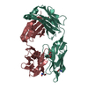
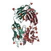
| Deposited unit | 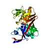
| ||||||||
|---|---|---|---|---|---|---|---|---|---|
| 1 | 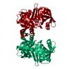
| ||||||||
| Unit cell |
|
-Components
| #1: Protein | Mass: 30054.732 Da / Num. of mol.: 1 / Fragment: Epimerase Source method: isolated from a genetically manipulated source Source: (gene. exp.) Corynebacterium glutamicum (strain ATCC 13032 / DSM 20300 / JCM 1318 / LMG 3730 / NCIMB 10025) (bacteria)Strain: ATCC 13032 / DSM 20300 / JCM 1318 / LMG 3730 / NCIMB 10025 Gene: dapF, Cgl1943, cg2129 / Production host: |
|---|---|
| #2: Chemical | ChemComp-API / 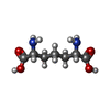  Type: L-peptide linking / Mass: 190.197 Da / Num. of mol.: 1 / Source method: obtained synthetically / Formula: C7H14N2O4 Type: L-peptide linking / Mass: 190.197 Da / Num. of mol.: 1 / Source method: obtained synthetically / Formula: C7H14N2O4 |
| #3: Water | ChemComp-HOH /  Mass: 18.015 Da / Num. of mol.: 68 / Source method: isolated from a natural source / Formula: H2O Mass: 18.015 Da / Num. of mol.: 68 / Source method: isolated from a natural source / Formula: H2O |
-Experimental details
-Experiment
| Experiment | Method: X-RAY DIFFRACTION / Number of used crystals: 1 |
|---|
- Sample preparation
| Crystal | Density Matthews: 5.3 Å3/Da / Density % sol: 76.77 % |
|---|---|
| Crystal grow | Temperature: 293 K / Method: vapor diffusion, hanging drop / pH: 9 / Details: Sodium citrate, CHES |
-Data collection
| Diffraction | Mean temperature: 100 K |
|---|---|
| Diffraction source | Source: SYNCHROTRON / Site: PAL/PLS  / Beamline: 7A (6B, 6C1) / Wavelength: 0.97934 Å / Beamline: 7A (6B, 6C1) / Wavelength: 0.97934 Å |
| Detector | Type: ADSC QUANTUM 270 / Detector: CCD / Date: Mar 15, 2015 |
| Radiation | Monochromator: Double Crystal Monochromator / Protocol: SINGLE WAVELENGTH / Monochromatic (M) / Laue (L): M / Scattering type: x-ray |
| Radiation wavelength | Wavelength: 0.97934 Å / Relative weight: 1 |
| Reflection | Resolution: 2.59→110.1 Å / Num. obs: 19071 / % possible obs: 97.4 % / Redundancy: 12 % / Rmerge(I) obs: 0.115 / Rsym value: 0.344 / Net I/σ(I): 21.1 |
| Reflection shell | Resolution: 2.6→2.64 Å / Redundancy: 4.5 % / Rmerge(I) obs: 0.344 / Mean I/σ(I) obs: 4 / % possible all: 93.4 |
- Processing
| Software |
| |||||||||||||||||||||||||||||||||||||||||||||||||||||||||||||||||||||||||||
|---|---|---|---|---|---|---|---|---|---|---|---|---|---|---|---|---|---|---|---|---|---|---|---|---|---|---|---|---|---|---|---|---|---|---|---|---|---|---|---|---|---|---|---|---|---|---|---|---|---|---|---|---|---|---|---|---|---|---|---|---|---|---|---|---|---|---|---|---|---|---|---|---|---|---|---|---|
| Refinement | Method to determine structure: MOLECULAR REPLACEMENT Starting model: 5H2Y Resolution: 2.59→110.09 Å / Cor.coef. Fo:Fc: 0.936 / Cor.coef. Fo:Fc free: 0.91 / SU B: 8.502 / SU ML: 0.161 / Cross valid method: THROUGHOUT / σ(F): 0 / ESU R: 0.231 / ESU R Free: 0.205 / Stereochemistry target values: MAXIMUM LIKELIHOOD Details: HYDROGENS HAVE BEEN ADDED IN THE RIDING POSITIONS U VALUES : REFINED INDIVIDUALLY
| |||||||||||||||||||||||||||||||||||||||||||||||||||||||||||||||||||||||||||
| Solvent computation | Ion probe radii: 0.8 Å / Shrinkage radii: 0.8 Å / VDW probe radii: 1.2 Å / Solvent model: MASK | |||||||||||||||||||||||||||||||||||||||||||||||||||||||||||||||||||||||||||
| Displacement parameters | Biso max: 113.57 Å2 / Biso mean: 34.446 Å2 / Biso min: 8.97 Å2
| |||||||||||||||||||||||||||||||||||||||||||||||||||||||||||||||||||||||||||
| Refinement step | Cycle: final / Resolution: 2.59→110.09 Å
| |||||||||||||||||||||||||||||||||||||||||||||||||||||||||||||||||||||||||||
| Refine LS restraints |
| |||||||||||||||||||||||||||||||||||||||||||||||||||||||||||||||||||||||||||
| LS refinement shell | Resolution: 2.594→2.662 Å / Total num. of bins used: 20
|
