Entry Database : PDB / ID : 5lyqTitle Crystal structure of the Retinoic Acid Receptor alpha in complex with a synthetic spiroketal agonist and a fragment of the TIF2 co-activator. HIS-LYS-ILE-LEU-HIS-ARG-LEU-LEU-GLN-ASP Retinoic acid receptor RXR-alpha Keywords / / / Function / homology Function Domain/homology Component
/ / / / / / / / / / / / / / / / / / / / / / / / / / / / / / / / / / / / / / / / / / / / / / / / / / / / / / / / / / / / / / / / / / / / / / / / / / / / / / / / / / / / / / / / / / / / / / / / / / / / / / / / / / / / / / / / / / / / / / / / / / / / / / / / / / / / / / / / / / / / / / / / / / / / / / / / / / / / / / / / / / / Biological species Homo sapiens (human)Method / / Resolution : 2.17 Å Authors Andrei, S.A. / Ottmann, C. Funding support Organization Grant number Country NWO ECHO 711011017
Journal : Angew. Chem. Int. Ed. Engl. / Year : 2017Title : Designed Spiroketal Protein Modulation.Authors : Scheepstra, M. / Andrei, S.A. / Unver, M.Y. / Hirsch, A.K.H. / Leysen, S. / Ottmann, C. / Brunsveld, L. / Milroy, L.G. History Deposition Sep 28, 2016 Deposition site / Processing site Revision 1.0 Apr 26, 2017 Provider / Type Revision 1.1 May 10, 2017 Group Revision 1.2 Sep 13, 2017 Group / Category / Item Revision 1.3 Aug 10, 2022 Group / Derived calculationsCategory database_2 / pdbx_struct_assembly ... database_2 / pdbx_struct_assembly / pdbx_struct_assembly_gen / pdbx_struct_assembly_prop / pdbx_struct_oper_list Item _database_2.pdbx_DOI / _database_2.pdbx_database_accession ... _database_2.pdbx_DOI / _database_2.pdbx_database_accession / _pdbx_struct_assembly.details / _pdbx_struct_assembly.method_details / _pdbx_struct_assembly.oligomeric_count / _pdbx_struct_assembly.oligomeric_details Revision 1.4 Jan 17, 2024 Group / Refinement descriptionCategory / chem_comp_bond / pdbx_initial_refinement_model
Show all Show less
 Movie
Movie Controller

 Yorodumi
 Open data
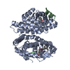
 Basic information
 Components
 Keywords
 Function and homology information
 Homo sapiens (human)
 X-RAY DIFFRACTION /
  Authors
 Netherlands, 1items
  Citation
 Structure visualization
 Downloads & links
 Other downloads
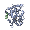
 PDBj
PDBj


 Assembly
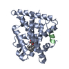

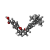
 Mass: 406.514 Da / Num. of mol.: 1 / Source method: obtained synthetically / Formula: C26H30O4
 Mass: 18.015 Da / Num. of mol.: 165 / Source method: isolated from a natural source / Formula: H2O
 Sample preparation
 Processing