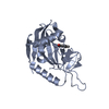
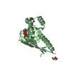
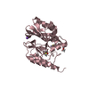
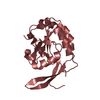
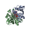
Journal: Mol Microbiol / Year: 2017 Title: Evolved distal tail carbohydrate binding modules of Lactobacillus phage J-1: a novel type of anti-receptor widespread among lactic acid bacteria phages. Authors: Maria-Eugenia Dieterle / Silvia Spinelli / Irina Sadovskaya / Mariana Piuri / Christian Cambillau / Abstract: Bacteriophage replication requires specific host-recognition. Some siphophages harbour a large complex, the baseplate, at the tip of their non-contractile tail. This baseplate holds receptor binding ...Bacteriophage replication requires specific host-recognition. Some siphophages harbour a large complex, the baseplate, at the tip of their non-contractile tail. This baseplate holds receptor binding proteins (RBPs) that can recognize the host cell-wall polysaccharide (CWPS) and specifically attach the phage to its host. While most phages possess a dedicated RBP, the phage J-1 that infects Lactobacillus casei seemed to lack one. It has been shown that the phage J-1 distal tail protein (Dit) plays a role in host recognition and that its sequence comprises two inserted modules compared with 'classical' Dits. The first insertion is similar to carbohydrate-binding modules (CBMs), whereas the second insertion remains undocumented. Here, we determined the structure of the second insertion and found it also similar to several CBMs. Expressed insertion CBM2, but not CBM1, binds to L. casei cells and neutralize phage attachment to the bacterial cell wall and the isolated and purified CWPS of L. casei BL23 prevents CBM2 attachment to the host. Electron microscopy single particle reconstruction of the J-1 virion baseplate revealed that CBM2 is projected at the periphery of Dit to optimally bind the CWPS receptor. Taken together, these results identify J-1 evolved Dit as the phage RBP.
Mass: 18.015 Da / Num. of mol.: 317 / Source method: isolated from a natural source / Formula: H2O
-
Experimental details
-
Experiment
Experiment
Method: X-RAY DIFFRACTION / Number of used crystals: 1
-
Sample preparation
Crystal
Density Matthews: 2.11 Å3/Da / Density % sol: 41.6 %
Crystal grow
Temperature: 293 K / Method: vapor diffusion, sitting drop Details: protein at 8 mg/mL in 0.8-1.2 M sodium citrate as precipitant with 10mM sodium borate at pH 8.9-9.7. PH range: 8.9-9.7
In the structure databanks used in Yorodumi, some data are registered as the other names, "COVID-19 virus" and "2019-nCoV". Here are the details of the virus and the list of structure data.
Jan 31, 2019. EMDB accession codes are about to change! (news from PDBe EMDB page)
EMDB accession codes are about to change! (news from PDBe EMDB page)
The allocation of 4 digits for EMDB accession codes will soon come to an end. Whilst these codes will remain in use, new EMDB accession codes will include an additional digit and will expand incrementally as the available range of codes is exhausted. The current 4-digit format prefixed with “EMD-” (i.e. EMD-XXXX) will advance to a 5-digit format (i.e. EMD-XXXXX), and so on. It is currently estimated that the 4-digit codes will be depleted around Spring 2019, at which point the 5-digit format will come into force.
The EM Navigator/Yorodumi systems omit the EMD- prefix.
Related info.:Q: What is EMD? / ID/Accession-code notation in Yorodumi/EM Navigator
Yorodumi is a browser for structure data from EMDB, PDB, SASBDB, etc.
This page is also the successor to EM Navigator detail page, and also detail information page/front-end page for Omokage search.
The word "yorodu" (or yorozu) is an old Japanese word meaning "ten thousand". "mi" (miru) is to see.
Related info.:EMDB / PDB / SASBDB / Comparison of 3 databanks / Yorodumi Search / Aug 31, 2016. New EM Navigator & Yorodumi / Yorodumi Papers / Jmol/JSmol / Function and homology information / Changes in new EM Navigator and Yorodumi
 Movie
Movie Controller

 Yorodumi
 Open data
 Basic information
 Components
 Keywords
 Function and homology information
 Lactobacillus phage J-1 (virus)
 X-RAY DIFFRACTION /
  Authors
 Citation

 Structure visualization
 Downloads & links
 Other downloads
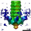


 PDBj
PDBj Assembly
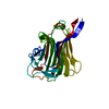


 Mass: 24.305 Da / Num. of mol.: 2 / Source method: obtained synthetically / Formula: Mg
 Mass: 18.015 Da / Num. of mol.: 317 / Source method: isolated from a natural source / Formula: H2O
 Sample preparation
 Processing