 Movie
Movie Controller

[English] 日本語
 Yorodumi
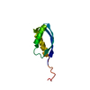
- PDB-5lxk: NMR structure of the C-terminal domain of the Bacteriophage T5 de... -
+ Open data

- Basic information
| Entry | Database: PDB / ID: 5lxk | ||||||
|---|---|---|---|---|---|---|---|
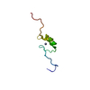
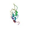
| Title | NMR structure of the C-terminal domain of the Bacteriophage T5 decoration protein pb10. | ||||||
 Components Components | Decoration protein | ||||||
 Keywords Keywords | VIRAL PROTEIN / Bacteriophage T5 Decoration Protein | ||||||
| Function / homology | : / Decoration protein pb10 N-terminal domain / viral capsid, decoration / Immunoglobulin domain / Ig-like domain profile. / Immunoglobulin-like domain / Immunoglobulin-like domain superfamily / Immunoglobulin-like fold / Decoration protein Function and homology information Function and homology information | ||||||
| Biological species |  Escherichia phage T5 (virus) Escherichia phage T5 (virus) | ||||||
| Method | SOLUTION NMR / simulated annealing | ||||||
 Authors Authors | Vernhes, E. / Gilquin, B. / Cuniasse, P. / Boulanger, P. / Zinn-Justin, S. | ||||||
| Funding support |  France, 1items France, 1items
| ||||||
 Citation Citation | Journal: Sci Rep / Year: 2017 Title: High affinity anchoring of the decoration protein pb10 onto the bacteriophage T5 capsid. Authors: Emeline Vernhes / Madalena Renouard / Bernard Gilquin / Philippe Cuniasse / Dominique Durand / Patrick England / Sylviane Hoos / Alexis Huet / James F Conway / Anatoly Glukhov / Vladimir ...Authors: Emeline Vernhes / Madalena Renouard / Bernard Gilquin / Philippe Cuniasse / Dominique Durand / Patrick England / Sylviane Hoos / Alexis Huet / James F Conway / Anatoly Glukhov / Vladimir Ksenzenko / Eric Jacquet / Naïma Nhiri / Sophie Zinn-Justin / Pascale Boulanger /   Abstract: Bacteriophage capsids constitute icosahedral shells of exceptional stability that protect the viral genome. Many capsids display on their surface decoration proteins whose structure and function ...Bacteriophage capsids constitute icosahedral shells of exceptional stability that protect the viral genome. Many capsids display on their surface decoration proteins whose structure and function remain largely unknown. The decoration protein pb10 of phage T5 binds at the centre of the 120 hexamers formed by the major capsid protein. Here we determined the 3D structure of pb10 and investigated its capsid-binding properties using NMR, SAXS, cryoEM and SPR. Pb10 consists of an α-helical capsid-binding domain and an Ig-like domain exposed to the solvent. It binds to the T5 capsid with a remarkably high affinity and its binding kinetics is characterized by a very slow dissociation rate. We propose that the conformational exchange events observed in the capsid-binding domain enable rearrangements upon binding that contribute to the quasi-irreversibility of the pb10-capsid interaction. Moreover we show that pb10 binding is a highly cooperative process, which favours immediate rebinding of newly dissociated pb10 to the 120 hexamers of the capsid protein. In extreme conditions, pb10 protects the phage from releasing its genome. We conclude that pb10 may function to reinforce the capsid thus favouring phage survival in harsh environments. | ||||||
| History |
|
- Structure visualization
| Structure viewer | Molecule: MolmilJmol/JSmol |
|---|
- Downloads & links
-Download
| PDBx/mmCIF format | 5lxk.cif.gz | 529.7 KB | Display | PDBx/mmCIF format |
|---|---|---|---|---|
| PDB format | pdb5lxk.ent.gz | 445.6 KB | Display | PDB format |
| PDBx/mmJSON format | 5lxk.json.gz | Tree view | PDBx/mmJSON format | |
| Others |  Other downloads Other downloads |
-Validation report
| Summary document | 5lxk_validation.pdf.gz | 410.1 KB | Display | wwPDB validaton report |
|---|---|---|---|---|
| Full document | 5lxk_full_validation.pdf.gz | 566.1 KB | Display | |
| Data in XML | 5lxk_validation.xml.gz | 32 KB | Display | |
| Data in CIF | 5lxk_validation.cif.gz | 56 KB | Display | |
| Arichive directory | https://data.pdbj.org/pub/pdb/validation_reports/lx/5lxkftp://data.pdbj.org/pub/pdb/validation_reports/lx/5lxk | HTTPS FTP |
-Related structure data
| Related structure data |  8419C 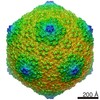 8423C  5lxlC 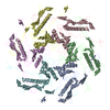 5tjtC C: citing same article ( |
|---|---|
| Similar structure data | |
| Other databases |
|


-Links
 PDBj
PDBj

- Assembly
| Deposited unit | 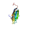
| |||||||||
|---|---|---|---|---|---|---|---|---|---|---|
| 1 |
| |||||||||
| NMR ensembles |
|
-Components
| #1: Protein | Mass: 10339.291 Da / Num. of mol.: 1 / Fragment: C-terminal domain, UNP residues 73-164 Source method: isolated from a genetically manipulated source Source: (gene. exp.) Escherichia phage T5 (virus) / Gene: N5, T5.151, T5p147 / Production host:  |
|---|
-Experimental details
-Experiment
| Experiment | Method: SOLUTION NMR | ||||||||||||||||||||||||||||||||||||||||||||||||||||||||||||||||||
|---|---|---|---|---|---|---|---|---|---|---|---|---|---|---|---|---|---|---|---|---|---|---|---|---|---|---|---|---|---|---|---|---|---|---|---|---|---|---|---|---|---|---|---|---|---|---|---|---|---|---|---|---|---|---|---|---|---|---|---|---|---|---|---|---|---|---|---|
| NMR experiment |
|
 HSQC
HSQC- Sample preparation
| Details | Type: solution Contents: 200 uM [U-99% 13C; U-99% 15N] c72, 90% H2O/10% D2O Label: 15N_C13_sample / Solvent system: 90% H2O/10% D2O |
|---|---|
| Sample | Conc.: 200 uM / Component: c72 / Isotopic labeling: [U-99% 13C; U-99% 15N] |
| Sample conditions | Ionic strength: 25 mM / Label: condition_1 / pH: 7.2 / Pressure: 1 atm / Temperature: 293 K |
-NMR measurement
| NMR spectrometer |
|
|---|
- Processing
| NMR software |
| ||||||||||||||||||
|---|---|---|---|---|---|---|---|---|---|---|---|---|---|---|---|---|---|---|---|
| Refinement | Method: simulated annealing / Software ordinal: 5 | ||||||||||||||||||
| NMR representative | Selection criteria: lowest energy | ||||||||||||||||||
| NMR ensemble | Conformer selection criteria: structures with the lowest energy Conformers calculated total number: 200 / Conformers submitted total number: 20 |
