 Movie
Movie Controller


+ Open data

- Basic information
| Entry | Database: PDB / ID: 5lsk | ||||||
|---|---|---|---|---|---|---|---|
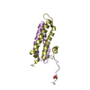
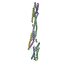
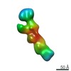
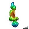
| Title | CRYSTAL STRUCTURE OF THE HUMAN KINETOCHORE MIS12-CENP-C COMPLEX | ||||||
 Components Components |
| ||||||
 Keywords Keywords | CELL CYCLE / ALPHA-HELICAL | ||||||
| Function / homology |  Function and homology information Function and homology informationskeletal muscle satellite cell proliferation / MIS12/MIND type complex / leucine zipper domain binding / spindle attachment to meiosis I kinetochore / attachment of spindle microtubules to kinetochore / centromeric DNA binding / kinetochore assembly / outer kinetochore / condensed chromosome, centromeric region / attachment of mitotic spindle microtubules to kinetochore ...skeletal muscle satellite cell proliferation / MIS12/MIND type complex / leucine zipper domain binding / spindle attachment to meiosis I kinetochore / attachment of spindle microtubules to kinetochore / centromeric DNA binding / kinetochore assembly / outer kinetochore / condensed chromosome, centromeric region / attachment of mitotic spindle microtubules to kinetochore / inner kinetochore / mitotic sister chromatid segregation / pericentric heterochromatin / Amplification of signal from unattached kinetochores via a MAD2 inhibitory signal / Deposition of new CENPA-containing nucleosomes at the centromere / Mitotic Prometaphase / EML4 and NUDC in mitotic spindle formation / Resolution of Sister Chromatid Cohesion / chromosome segregation / RHO GTPases Activate Formins / kinetochore / fibrillar center / spindle pole / azurophil granule lumen / Separation of Sister Chromatids / mitotic cell cycle / midbody / transcription regulator complex / transcription by RNA polymerase II / transcription coactivator activity / nuclear speck / nuclear body / cell division / Neutrophil degranulation / nucleolus / Golgi apparatus / DNA binding / extracellular region / nucleoplasm / identical protein binding / nucleus / cytosol Similarity search - Function | ||||||
| Biological species |  Homo sapiens (human) Homo sapiens (human) | ||||||
| Method |  X-RAY DIFFRACTION / SYNCHROTRON / SAD / Resolution: 3.502 Å X-RAY DIFFRACTION / SYNCHROTRON / SAD / Resolution: 3.502 Å | ||||||
 Authors Authors | Vetter, I.R. / Petrovic, A. / Keller, J. / Liu, Y. | ||||||
 Citation Citation | Journal: Cell / Year: 2016 Title: Structure of the MIS12 Complex and Molecular Basis of Its Interaction with CENP-C at Human Kinetochores. Authors: Petrovic, A. / Keller, J. / Liu, Y. / Overlack, K. / John, J. / Dimitrova, Y.N. / Jenni, S. / van Gerwen, S. / Stege, P. / Wohlgemuth, S. / Rombaut, P. / Herzog, F. / Harrison, S.C. / ...Authors: Petrovic, A. / Keller, J. / Liu, Y. / Overlack, K. / John, J. / Dimitrova, Y.N. / Jenni, S. / van Gerwen, S. / Stege, P. / Wohlgemuth, S. / Rombaut, P. / Herzog, F. / Harrison, S.C. / Vetter, I.R. / Musacchio, A. | ||||||
| History |
|
- Structure visualization
| Structure viewer | Molecule: MolmilJmol/JSmol |
|---|
- Downloads & links
-Download
| PDBx/mmCIF format | 5lsk.cif.gz | 171.9 KB | Display | PDBx/mmCIF format |
|---|---|---|---|---|
| PDB format | pdb5lsk.ent.gz | 131.8 KB | Display | PDB format |
| PDBx/mmJSON format | 5lsk.json.gz | Tree view | PDBx/mmJSON format | |
| Others |  Other downloads Other downloads |
-Validation report
| Arichive directory | https://data.pdbj.org/pub/pdb/validation_reports/ls/5lskftp://data.pdbj.org/pub/pdb/validation_reports/ls/5lsk | HTTPS FTP |
|---|
-Related structure data


-Links
 PDBj
PDBj


- Assembly
| Deposited unit | 
| ||||||||
|---|---|---|---|---|---|---|---|---|---|
| 1 |
| ||||||||
| Unit cell |
|
-Components
-Protein , 3 types, 3 molecules ABP
| #1: Protein | Mass: 24170.922 Da / Num. of mol.: 1 Source method: isolated from a genetically manipulated source Source: (gene. exp.) Homo sapiens (human) / Gene: MIS12 / Plasmid: pST39 / Production host:  |
|---|---|
| #2: Protein | Mass: 20522.359 Da / Num. of mol.: 1 / Fragment: UNP residues 31-205 Source method: isolated from a genetically manipulated source Source: (gene. exp.) Homo sapiens (human) / Gene: PMF1 / Plasmid: pST39 / Production host: |
| #5: Protein | Mass: 8501.529 Da / Num. of mol.: 1 Source method: isolated from a genetically manipulated source Source: (gene. exp.) Homo sapiens (human) / Gene: CENPC, CENPC1, ICEN7 / Production host: |
-Kinetochore-associated protein ... , 2 types, 2 molecules DN
| #3: Protein | Mass: 33848.191 Da / Num. of mol.: 1 / Fragment: UNP residues 68-356 Source method: isolated from a genetically manipulated source Source: (gene. exp.) Homo sapiens (human) / Gene: DSN1, C20orf172, MIS13 / Plasmid: pST39 / Production host: |
|---|---|
| #4: Protein | Mass: 23307.619 Da / Num. of mol.: 1 Source method: isolated from a genetically manipulated source Source: (gene. exp.) Homo sapiens (human) / Gene: NSL1, C1orf48, DC31, DC8, MIS14 / Production host: |
-Non-polymers , 1 types, 6 molecules 
| #6: Water | ChemComp-HOH / Mass: 18.015 Da / Num. of mol.: 6 / Source method: isolated from a natural source / Formula: H2O |
|---|
-Details
| Has protein modification | Y |
|---|
-Experimental details
-Experiment
| Experiment | Method: X-RAY DIFFRACTION / Number of used crystals: 1 |
|---|
- Sample preparation
| Crystal | Density Matthews: 3.9 Å3/Da / Density % sol: 67.9 % |
|---|---|
| Crystal grow | Temperature: 277 K / Method: vapor diffusion / Details: 6-12% PEG6000 / PH range: 6-8 |
-Data collection
| Diffraction | Mean temperature: 100 K | ||||||||||||||||||||||||||||||||||||||||||||||||||||||||||||||||||||||||||||||||||||||||||||||||||||||||||||||||||||||||||||||
|---|---|---|---|---|---|---|---|---|---|---|---|---|---|---|---|---|---|---|---|---|---|---|---|---|---|---|---|---|---|---|---|---|---|---|---|---|---|---|---|---|---|---|---|---|---|---|---|---|---|---|---|---|---|---|---|---|---|---|---|---|---|---|---|---|---|---|---|---|---|---|---|---|---|---|---|---|---|---|---|---|---|---|---|---|---|---|---|---|---|---|---|---|---|---|---|---|---|---|---|---|---|---|---|---|---|---|---|---|---|---|---|---|---|---|---|---|---|---|---|---|---|---|---|---|---|---|---|
| Diffraction source | Source: SYNCHROTRON / Site: SLS  / Beamline: X10SA / Wavelength: 0.97863 Å / Beamline: X10SA / Wavelength: 0.97863 Å | ||||||||||||||||||||||||||||||||||||||||||||||||||||||||||||||||||||||||||||||||||||||||||||||||||||||||||||||||||||||||||||||
| Detector | Type: DECTRIS PILATUS3 S 6M / Detector: PIXEL / Date: Mar 19, 2015 | ||||||||||||||||||||||||||||||||||||||||||||||||||||||||||||||||||||||||||||||||||||||||||||||||||||||||||||||||||||||||||||||
| Radiation | Protocol: SINGLE WAVELENGTH / Monochromatic (M) / Laue (L): M / Scattering type: x-ray | ||||||||||||||||||||||||||||||||||||||||||||||||||||||||||||||||||||||||||||||||||||||||||||||||||||||||||||||||||||||||||||||
| Radiation wavelength | Wavelength: 0.97863 Å / Relative weight: 1 | ||||||||||||||||||||||||||||||||||||||||||||||||||||||||||||||||||||||||||||||||||||||||||||||||||||||||||||||||||||||||||||||
| Reflection | Resolution: 3.5→19.742 Å / Num. obs: 16932 / % possible obs: 99 % / Observed criterion σ(I): -3 / Redundancy: 6.76 % / Biso Wilson estimate: 98.247 Å2 / Rmerge(I) obs: 0.181 / Net I/σ(I): 7.16 | ||||||||||||||||||||||||||||||||||||||||||||||||||||||||||||||||||||||||||||||||||||||||||||||||||||||||||||||||||||||||||||||
| Reflection shell |
|
- Processing
| Software |
| ||||||||||||||||||||||||||||||||||||||||||
|---|---|---|---|---|---|---|---|---|---|---|---|---|---|---|---|---|---|---|---|---|---|---|---|---|---|---|---|---|---|---|---|---|---|---|---|---|---|---|---|---|---|---|---|
| Refinement | Method to determine structure: SAD / Resolution: 3.502→19.742 Å / SU ML: 0.5 / Cross valid method: FREE R-VALUE / σ(F): 1.37 / Phase error: 30.84
| ||||||||||||||||||||||||||||||||||||||||||
| Solvent computation | Shrinkage radii: 0.9 Å / VDW probe radii: 1.11 Å | ||||||||||||||||||||||||||||||||||||||||||
| Displacement parameters | Biso max: 167.51 Å2 / Biso mean: 74.2897 Å2 / Biso min: 22.94 Å2 | ||||||||||||||||||||||||||||||||||||||||||
| Refinement step | Cycle: LAST / Resolution: 3.502→19.742 Å
| ||||||||||||||||||||||||||||||||||||||||||
| Refine LS restraints |
| ||||||||||||||||||||||||||||||||||||||||||
| LS refinement shell | Refine-ID: X-RAY DIFFRACTION / Total num. of bins used: 5
|
