[English] 日本語
Yorodumi
- PDB-5lrt: Structure of the Deamidase-Depupylase Dop of the Prokaryotic Ubiq... -

+
Open data


ID or keywords:

Loading...

-
Basic information

Entry
Database: PDB / ID: 5lrt
Title
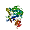
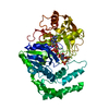
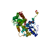
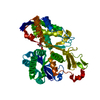
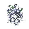
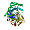
Structure of the Deamidase-Depupylase Dop of the Prokaryotic Ubiquitin-like Modification Pathway in Complex with ADP and Phosphate
ComponentsDepupylase
KeywordsHYDROLASE / Complex
Function / homology
Function and homology information


protein pupylation / hydrolase activity, acting on carbon-nitrogen (but not peptide) bonds, in linear amides / Hydrolases; Acting on peptide bonds (peptidases) / proteasomal protein catabolic process / modification-dependent protein catabolic process / peptidase activity / ATP binding / metal ion binding
Similarity search - Function
Pup deamidase / Pup ligase/deamidase / Pup-ligase protein
Similarity search - Domain/homology
ADENOSINE-5'-DIPHOSPHATE / DI(HYDROXYETHYL)ETHER / TRIETHYLENE GLYCOL / PHOSPHATE ION / Depupylase
Similarity search - Component
Biological speciesAcidothermus cellulolyticus (bacteria)
MethodX-RAY DIFFRACTION / SYNCHROTRON / MOLECULAR REPLACEMENT / molecular replacement / Resolution: 1.85 Å
AuthorsBolten, M. / Vahlensieck, C. / Lipp, C. / Leibundgut, M. / Ban, N. / Weber-Ban, E.
CitationJournal: J. Biol. Chem. / Year: 2017
Title: Depupylase Dop Requires Inorganic Phosphate in the Active Site for Catalysis.
Authors: Bolten, M. / Vahlensieck, C. / Lipp, C. / Leibundgut, M. / Ban, N. / Weber-Ban, E.
History
DepositionAug 19, 2016Deposition site: PDBE / Processing site: PDBE
Revision 1.0Feb 1, 2017Provider: repository / Type: Initial release
Revision 1.1Feb 8, 2017Group: Database references
Revision 1.2Mar 22, 2017Group: Database references
Revision 1.3Jan 17, 2024Group: Data collection / Database references ...Data collection / Database references / Derived calculations / Refinement description
Category: chem_comp_atom / chem_comp_bond ...chem_comp_atom / chem_comp_bond / database_2 / pdbx_initial_refinement_model / pdbx_struct_conn_angle / struct_conn
Item: _database_2.pdbx_DOI / _database_2.pdbx_database_accession ..._database_2.pdbx_DOI / _database_2.pdbx_database_accession / _pdbx_struct_conn_angle.ptnr1_auth_comp_id / _pdbx_struct_conn_angle.ptnr1_auth_seq_id / _pdbx_struct_conn_angle.ptnr1_label_asym_id / _pdbx_struct_conn_angle.ptnr1_label_atom_id / _pdbx_struct_conn_angle.ptnr1_label_comp_id / _pdbx_struct_conn_angle.ptnr1_label_seq_id / _pdbx_struct_conn_angle.ptnr2_auth_seq_id / _pdbx_struct_conn_angle.ptnr2_label_asym_id / _pdbx_struct_conn_angle.ptnr3_auth_comp_id / _pdbx_struct_conn_angle.ptnr3_auth_seq_id / _pdbx_struct_conn_angle.ptnr3_label_asym_id / _pdbx_struct_conn_angle.ptnr3_label_atom_id / _pdbx_struct_conn_angle.ptnr3_label_comp_id / _pdbx_struct_conn_angle.ptnr3_label_seq_id / _pdbx_struct_conn_angle.value / _struct_conn.pdbx_dist_value / _struct_conn.ptnr1_label_atom_id / _struct_conn.ptnr2_auth_comp_id / _struct_conn.ptnr2_auth_seq_id / _struct_conn.ptnr2_label_asym_id / _struct_conn.ptnr2_label_atom_id / _struct_conn.ptnr2_label_comp_id

-
Structure visualization

Structure viewerMolecule:
MolmilJmol/JSmol

Downloads & links

-
Assembly

Deposited unit
A: Depupylase
hetero molecules


Theoretical massNumber of molelcules
Total (without water)58,62012
Polymers57,2881
Non-polymers1,33111
Water4,035224
1


  • Idetical with deposited unit
  • defined by author&software
TypeNameSymmetry operationNumber
identity operation1_555x,y,z1
Buried area3260 Å2
ΔGint-54 kcal/mol
Surface area18200 Å2
MethodPISA
Unit cell
Length a, b, c (Å)72.398, 72.398, 215.250
Angle α, β, γ (deg.)90.000, 90.000, 120.000
Int Tables number152
Space group name H-MP3121

-
Components

-
Protein , 1 types, 1 molecules A

#1: Protein Depupylase


Mass: 57288.332 Da / Num. of mol.: 1
Source method: isolated from a genetically manipulated source
Source: (gene. exp.) Acidothermus cellulolyticus (strain ATCC 43068 / 11B) (bacteria)
Gene: dop, Acel_1186 / Plasmid: pET21 / Production host: Escherichia coli BL21(DE3) (bacteria)
References: UniProt: A0LU48, Hydrolases; Acting on peptide bonds (peptidases)

-
Non-polymers , 8 types, 235 molecules

#2: Chemical ChemComp-ADP / ADENOSINE-5'-DIPHOSPHATE


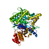
Mass: 427.201 Da / Num. of mol.: 1 / Source method: obtained synthetically / Formula: C10H15N5O10P2 / Comment: ADP, energy-carrying molecule*YM
#3: Chemical
ChemComp-MG / MAGNESIUM ION


Mass: 24.305 Da / Num. of mol.: 4 / Source method: obtained synthetically / Formula: Mg
#4: Chemical ChemComp-PO4 / PHOSPHATE ION


Mass: 94.971 Da / Num. of mol.: 1 / Source method: obtained synthetically / Formula: PO4
#5: Chemical ChemComp-P6G / HEXAETHYLENE GLYCOL / POLYETHYLENE GLYCOL PEG400


Mass: 282.331 Da / Num. of mol.: 1 / Source method: obtained synthetically / Formula: C12H26O7 / Comment: precipitant*YM
#6: Chemical ChemComp-PGE / TRIETHYLENE GLYCOL


Mass: 150.173 Da / Num. of mol.: 2 / Source method: obtained synthetically / Formula: C6H14O4
#7: Chemical ChemComp-PEG / DI(HYDROXYETHYL)ETHER


Mass: 106.120 Da / Num. of mol.: 1 / Source method: obtained synthetically / Formula: C4H10O3
#8: Chemical ChemComp-NA / SODIUM ION


Mass: 22.990 Da / Num. of mol.: 1 / Source method: obtained synthetically / Formula: Na
#9: Water ChemComp-HOH / water


Mass: 18.015 Da / Num. of mol.: 224 / Source method: isolated from a natural source / Formula: H2O

-
Experimental details

-
Experiment

ExperimentMethod: X-RAY DIFFRACTION / Number of used crystals: 1

-
Sample preparation

CrystalDensity Matthews: 2.84 Å3/Da / Density % sol: 56.73 %
Crystal growTemperature: 293 K / Method: vapor diffusion, sitting drop
Details: 18-23% (w/v) PEG 3350, 100mM bis-tris propane pH 8.25-9.0 at 20 degC, 200mM KSCN, 20mM MgCl2 and 5mM ATP
PH range: 8.25 - 9

-
Data collection

DiffractionMean temperature: 100 K
Diffraction sourceSource: SYNCHROTRON / Site: SLS / Beamline: X06SA / Wavelength: 0.99998 Å
DetectorType: DECTRIS PILATUS 6M / Detector: PIXEL / Date: Aug 22, 2013
RadiationProtocol: SINGLE WAVELENGTH / Monochromatic (M) / Laue (L): M / Scattering type: x-ray
Radiation wavelengthWavelength: 0.99998 Å / Relative weight: 1
ReflectionResolution: 1.85→36.2 Å / Num. obs: 56972 / % possible obs: 100 % / Redundancy: 8.9 % / Biso Wilson estimate: 39.17 Å2 / CC1/2: 1 / Rmerge(I) obs: 0.044 / Rpim(I) all: 0.016 / Rrim(I) all: 0.047 / Net I/σ(I): 24.3 / Num. measured all: 506782
Reflection shell
Resolution (Å)Redundancy (%)Rmerge(I) obsCC1/2Diffraction-ID% possible all
1.85-1.8991.4980.6151100
9.06-36.28.40.0221198.9

-
Phasing

PhasingMethod: molecular replacement
Phasing MR
Highest resolutionLowest resolution
Rotation7.27 Å36.2 Å
Translation7.27 Å36.2 Å

-
Processing

Software
NameVersionClassification
Aimless0.5.23data scaling
PHASER2.6.0phasing
PHENIX1.10_2155refinement
PDB_EXTRACT3.2data extraction
XDSVERSION March 30, 2013data reduction
RefinementMethod to determine structure: MOLECULAR REPLACEMENT
Starting model: PDB 4b0r
Resolution: 1.85→36.199 Å / SU ML: 0.23 / Cross valid method: FREE R-VALUE / σ(F): 1.34 / Phase error: 18.34
RfactorNum. reflection% reflection
Rfree0.1977 2880 5.06 %
Rwork0.1665 --
obs0.168 56895 99.98 %
Solvent computationShrinkage radii: 0.9 Å / VDW probe radii: 1.11 Å
Displacement parametersBiso max: 108.57 Å2 / Biso mean: 49.5952 Å2 / Biso min: 25.1 Å2
Refinement stepCycle: final / Resolution: 1.85→36.199 Å
ProteinNucleic acidLigandSolventTotal
Num. atoms3636 0 83 224 3943
Biso mean--52.69 53.07 -
Num. residues----464
Refine LS restraints
Refine-IDTypeDev idealNumber
X-RAY DIFFRACTIONf_bond_d0.0113849
X-RAY DIFFRACTIONf_angle_d1.1275229
X-RAY DIFFRACTIONf_chiral_restr0.066574
X-RAY DIFFRACTIONf_plane_restr0.008684
X-RAY DIFFRACTIONf_dihedral_angle_d14.9722336
LS refinement shell

Refine-ID: X-RAY DIFFRACTION / Total num. of bins used: 21 / % reflection obs: 100 %

Resolution (Å)Rfactor RfreeNum. reflection RfreeRfactor RworkNum. reflection RworkNum. reflection all
1.8501-1.88040.36941250.318524972622
1.8804-1.91280.31441500.303625522702
1.9128-1.94760.26791270.250325332660
1.9476-1.98510.24311610.229524942655
1.9851-2.02560.26931290.202225742703
2.0256-2.06960.19681390.194225052644
2.0696-2.11780.21411370.190425322669
2.1178-2.17070.18681300.176125402670
2.1707-2.22940.20741400.17225502690
2.2294-2.2950.20711360.165425442680
2.295-2.36910.22451270.166425692696
2.3691-2.45370.19391220.161425582680
2.4537-2.55190.16031440.159525652709
2.5519-2.6680.21381320.157525852717
2.668-2.80870.22331610.167625342695
2.8087-2.98450.21961400.169725792719
2.9845-3.21490.21221370.175225902727
3.2149-3.53810.20511300.167626012731
3.5381-4.04950.17641540.156126212775
4.0495-5.09970.17341250.135426722797
5.0997-36.20590.1791340.169728202954
Refinement TLS params.

Method: refined / Refine-ID: X-RAY DIFFRACTION

IDL112)L122)L132)L222)L232)L332)S11 (Å °)S12 (Å °)S13 (Å °)S21 (Å °)S22 (Å °)S23 (Å °)S31 (Å °)S32 (Å °)S33 (Å °)T112)T122)T132)T222)T232)T332)Origin x (Å)Origin y (Å)Origin z (Å)
11.9119-0.28411.76653.19570.54861.9218-0.19250.1851-0.02660.17430.3620.12750.0562-0.29130.02470.28790.01410.0630.3272-0.05260.3567-9.503466.652825.1164
20.2921-0.40340.00530.60950.17930.604-0.13750.11530.50370.62760.18630.3176-0.4088-0.23420.00020.56580.10140.07020.5179-0.06940.4871-12.007176.529733.7519
31.43190.91370.90823.06880.28621.89220.0130.04520.05620.31580.0786-0.017-0.22870.0668-0.00020.2873-0.01190.02310.3194-0.03140.3055-3.763171.123827.7499
40.2502-0.0337-0.00490.104-0.10320.11560.0636-0.82390.30010.8767-0.01520.0168-0.2707-0.35980.00050.68690.02410.11920.6638-0.10220.469-14.708766.79640.0921
51.19720.27091.0262.25890.99681.6513-0.0920.5741-0.1826-0.26740.13290.1976-0.1492-0.13330.00750.31-0.02730.02230.5143-0.09120.3221-11.865861.084511.6646
60.0156-0.0199-0.00390.02630.00940.0194-0.04680.16360.1036-0.11820.15110.4856-0.1012-0.0552-0.00130.7211-0.02460.03670.7214-0.07961.0215-21.217469.497812.2116
71.26380.08290.51021.42020.55761.3894-0.18810.69270.0205-0.44210.2824-0.2469-0.41610.33570.01280.4222-0.09590.06340.586-0.04590.3398-3.168268.61199.371
80.3144-0.06710.11570.4362-0.04050.2712-0.04760.8919-0.5384-0.3130.1522-0.11090.2240.26450.03750.4449-0.05810.02170.7694-0.28820.5026-12.363151.61923.0114
90.7091-0.27870.67861.020.37941.13080.01450.8143-0.2767-0.60960.1126-0.35090.01430.6396-0.14920.43440.04230.22450.9348-0.57940.60944.979254.32253.7142
100.93070.4901-0.60441.5091-0.38091.93270.07280.0148-0.45610.3403-0.008-0.21810.3750.0376-0.00020.33390.0364-0.05120.3326-0.05330.5201-5.781250.006428.109
112.01340.83570.63020.85131.00211.4909-0.32010.27510.708-0.16510.2673-0.2519-0.47760.3667-0.00040.4572-0.1381-0.00460.44020.00320.51046.407787.921325.2276
Refinement TLS group
IDRefine-IDRefine TLS-IDSelection detailsAuth asym-IDAuth seq-ID
1X-RAY DIFFRACTION1(chain A and resid 1:20)A1 - 20
2X-RAY DIFFRACTION2(chain A and resid 21:42)A21 - 43
3X-RAY DIFFRACTION3(chain A and resid 79:130)A79 - 130
4X-RAY DIFFRACTION4(chain A and resid 131:141)A131 - 141
5X-RAY DIFFRACTION5(chain A and resid 142:215)A142 - 215
6X-RAY DIFFRACTION6(chain A and resid 216:220)A216 - 220
7X-RAY DIFFRACTION7(chain A and resid 221:273)A221 - 273
8X-RAY DIFFRACTION8(chain A and resid 274:299)A274 - 299
9X-RAY DIFFRACTION9(chain A and resid 300:353)A300 - 353
10X-RAY DIFFRACTION10(chain A and resid 354:425)A354 - 425
11X-RAY DIFFRACTION11(chain A and resid 426:500)A426 - 500

+
About Yorodumi

-
News

-
Feb 9, 2022. New format data for meta-information of EMDB entries

New format data for meta-information of EMDB entries

  • Version 3 of the EMDB header file is now the official format.
  • The previous official version 1.9 will be removed from the archive.

Related info.:EMDB header

External links:wwPDB to switch to version 3 of the EMDB data model

-
Aug 12, 2020. Covid-19 info

Covid-19 info

URL: https://pdbjlvh1.pdbj.org/emnavi/covid19.php

New page: Covid-19 featured information page in EM Navigator.

Related info.:Covid-19 info / Mar 5, 2020. Novel coronavirus structure data

+
Mar 5, 2020. Novel coronavirus structure data

Novel coronavirus structure data

Related info.:Yorodumi Speices / Aug 12, 2020. Covid-19 info

External links:COVID-19 featured content - PDBj / Molecule of the Month (242):Coronavirus Proteases

+
Jan 31, 2019. EMDB accession codes are about to change! (news from PDBe EMDB page)

EMDB accession codes are about to change! (news from PDBe EMDB page)

  • The allocation of 4 digits for EMDB accession codes will soon come to an end. Whilst these codes will remain in use, new EMDB accession codes will include an additional digit and will expand incrementally as the available range of codes is exhausted. The current 4-digit format prefixed with “EMD-” (i.e. EMD-XXXX) will advance to a 5-digit format (i.e. EMD-XXXXX), and so on. It is currently estimated that the 4-digit codes will be depleted around Spring 2019, at which point the 5-digit format will come into force.
  • The EM Navigator/Yorodumi systems omit the EMD- prefix.

Related info.:Q: What is EMD? / ID/Accession-code notation in Yorodumi/EM Navigator

External links:EMDB Accession Codes are Changing Soon! / Contact to PDBj

+
Jul 12, 2017. Major update of PDB

Major update of PDB

  • wwPDB released updated PDB data conforming to the new PDBx/mmCIF dictionary.
  • This is a major update changing the version number from 4 to 5, and with Remediation, in which all the entries are updated.
  • In this update, many items about electron microscopy experimental information are reorganized (e.g. em_software).
  • Now, EM Navigator and Yorodumi are based on the updated data.

External links:wwPDB Remediation / Enriched Model Files Conforming to OneDep Data Standards Now Available in the PDB FTP Archive

-
Yorodumi

Thousand views of thousand structures

  • Yorodumi is a browser for structure data from EMDB, PDB, SASBDB, etc.
  • This page is also the successor to EM Navigator detail page, and also detail information page/front-end page for Omokage search.
  • The word "yorodu" (or yorozu) is an old Japanese word meaning "ten thousand". "mi" (miru) is to see.

Related info.:EMDB / PDB / SASBDB / Comparison of 3 databanks / Yorodumi Search / Aug 31, 2016. New EM Navigator & Yorodumi / Yorodumi Papers / Jmol/JSmol / Function and homology information / Changes in new EM Navigator and Yorodumi

Read more