Protocol: SINGLE WAVELENGTH / Monochromatic (M) / Laue (L): M / Scattering type: x-ray
Radiation wavelength
Wavelength: 0.975 Å / Relative weight: 1
Reflection twin
Crystal-ID
ID
Operator
Domain-ID
Fraction
1
1
H, K, L
1
0.425
1
1
-h,-k,l
2
0.575
Reflection
Resolution: 2.2→33.91 Å / Num. all: 497070 / Num. obs: 161184 / % possible obs: 99 % / Redundancy: 3.1 % / Rmerge(I) obs: 0.097 / Net I/σ(I): 10.4
Reflection shell
Resolution: 2.2→2.24 Å / Rmerge(I) obs: 0.41
-
Processing
Software
Name
Version
Classification
REFMAC
5.8.0103
refinement
XDS
datareduction
Aimless
datascaling
MOLREP
phasing
Refinement
Resolution: 2.2→31.92 Å / Cor.coef. Fo:Fc: 0.947 / Cor.coef. Fo:Fc free: 0.911 / SU B: 3.857 / SU ML: 0.104 / Cross valid method: THROUGHOUT / ESU R: 0.042 / ESU R Free: 0.037 / Details: HYDROGENS HAVE BEEN ADDED IN THE RIDING POSITIONS
Rfactor
Num. reflection
% reflection
Selection details
Rfree
0.20461
8412
4.9 %
RANDOM
Rwork
0.15011
-
-
-
obs
0.15282
153197
98.28 %
-
Solvent computation
Ion probe radii: 0.8 Å / Shrinkage radii: 0.8 Å / VDW probe radii: 1.2 Å
 Movie
Movie Controller

 Yorodumi
 Open data
 Basic information
 Components
 Keywords
 Function and homology information
 X-RAY DIFFRACTION /
  Authors
 Citation
 Structure visualization
 Downloads & links
 Other downloads
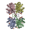


 PDBj
PDBj

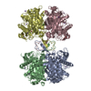
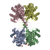
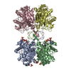
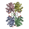
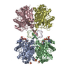
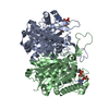
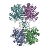
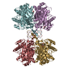
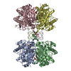
 Assembly
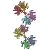

 Mass: 92.094 Da / Num. of mol.: 8 / Source method: obtained synthetically / Formula: C3H8O3
 Mass: 18.015 Da / Num. of mol.: 878 / Source method: isolated from a natural source / Formula: H2O
 Sample preparation
 / Beamline: I04 / Wavelength: 0.975 Å
 Processing