| Entry | Database: PDB / ID: 5loz
|
|---|
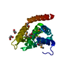
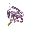
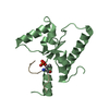
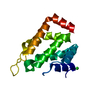
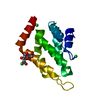
| Title | STRUCTURE OF YEAST ENT1 ENTH DOMAIN |
|---|
 Components Components | Epsin-1 |
|---|
 Keywords Keywords | UBIQUITIN-BINDING DOMAIN / ALPHA-ALPHA SUPERHELIX / UBIQUITIN RECEPTOR / ENDOCYTOSIS ADAPTOR / UBIQUITIN / CLATHRIN / LIPID / EPS15 |
|---|
| Function / homology |  Function and homology information Function and homology information
Cargo recognition for clathrin-mediated endocytosis / actin cortical patch assembly / clathrin vesicle coat / cellular bud tip / cellular bud neck / mating projection tip / K63-linked polyubiquitin modification-dependent protein binding / clathrin binding / actin filament organization / ubiquitin binding ...Cargo recognition for clathrin-mediated endocytosis / actin cortical patch assembly / clathrin vesicle coat / cellular bud tip / cellular bud neck / mating projection tip / K63-linked polyubiquitin modification-dependent protein binding / clathrin binding / actin filament organization / ubiquitin binding / phospholipid binding / endocytosis / early endosome / endosome / plasma membrane / cytoplasmSimilarity search - Function ENTH domain / Epsin N-terminal homology (ENTH) domain / ENTH domain profile. / ENTH domain / Serine Threonine Protein Phosphatase 5, Tetratricopeptide repeat - #90 / ENTH/VHS / Ubiquitin-interacting motif. / Ubiquitin interacting motif / Ubiquitin-interacting motif (UIM) domain profile. / Serine Threonine Protein Phosphatase 5, Tetratricopeptide repeat ...ENTH domain / Epsin N-terminal homology (ENTH) domain / ENTH domain profile. / ENTH domain / Serine Threonine Protein Phosphatase 5, Tetratricopeptide repeat - #90 / ENTH/VHS / Ubiquitin-interacting motif. / Ubiquitin interacting motif / Ubiquitin-interacting motif (UIM) domain profile. / Serine Threonine Protein Phosphatase 5, Tetratricopeptide repeat / Alpha Horseshoe / Mainly AlphaSimilarity search - Domain/homology |
|---|
| Biological species |   Saccharomyces cerevisiae (brewer's yeast) Saccharomyces cerevisiae (brewer's yeast) |
|---|
| Method |  X-RAY DIFFRACTION / SYNCHROTRON / MOLECULAR REPLACEMENT / Resolution: 1.95 Å X-RAY DIFFRACTION / SYNCHROTRON / MOLECULAR REPLACEMENT / Resolution: 1.95 Å |
|---|
 Authors Authors | Tanner, N. / Prag, G. |
|---|
| Funding support |  Israel, 1items Israel, 1items | Organization | Grant number | Country |
|---|
| Israel Science Foundation | 464/11 | Israel |
|
|---|
 Citation Citation | Journal: Nat.Methods / Year: 2016Title: A bacterial genetic selection system for ubiquitylation cascade discovery. Authors: Levin-Kravets, O. / Tanner, N. / Shohat, N. / Attali, I. / Keren-Kaplan, T. / Shusterman, A. / Artzi, S. / Varvak, A. / Reshef, Y. / Shi, X. / Zucker, O. / Baram, T. / Katina, C. / Pilzer, I. ...Authors: Levin-Kravets, O. / Tanner, N. / Shohat, N. / Attali, I. / Keren-Kaplan, T. / Shusterman, A. / Artzi, S. / Varvak, A. / Reshef, Y. / Shi, X. / Zucker, O. / Baram, T. / Katina, C. / Pilzer, I. / Ben-Aroya, S. / Prag, G. #1: Journal: Acta Crystallogr.,Sect.F / Year: 2012Title: Purification And Crystallization Of Yeast Ent1 Enth Domain. Authors: Levin Kravets, O. / Tanner, N. / Shohat, N. / Artzi, S. / Varvak, A. / Reshef, Y. / Attali, I. / Katina, C. / Keren Kaplan, T. / Prag, G. #2: Journal: Embo J. / Year: 2012Title: Synthetic Biology Approach To Reconstituting The Ubiquitylation Cascade In Bacteria. Authors: Keren-Kaplan, T. / Attali, I. / Motamedchaboki, K. / Davis, B.A. / Tanner, N. / Reshef, Y. / Laudon, E. / Kolot, M. / Levin-Kravets, O. / Kleifeld, O. / Glickman, M. / Horazdovsky, B.F. / Wolf, D.A. / Prag, G. |
|---|
| History | | Deposition | Aug 11, 2016 | Deposition site: PDBE / Processing site: PDBE |
|---|
| Revision 1.0 | Oct 5, 2016 | Provider: repository / Type: Initial release |
|---|
| Revision 1.1 | Oct 19, 2016 | Group: Database references |
|---|
| Revision 1.2 | Nov 9, 2016 | Group: Database references |
|---|
| Revision 1.3 | May 8, 2024 | Group: Data collection / Database references / Category: chem_comp_atom / chem_comp_bond / database_2
Item: _database_2.pdbx_DOI / _database_2.pdbx_database_accession |
|---|
|
|---|
 Movie
Movie Controller


 Open data
 Basic information
 Structure visualization
 Downloads & links
 Other downloads


 PDBj
PDBj


 Assembly
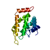

 Mass: 18.015 Da / Num. of mol.: 97 / Source method: isolated from a natural source / Formula: H2O
 Sample preparation
 / Beamline: ID23-1 / Wavelength: 0.979 Å
 Processing