 Movie
Movie Controller

[English] 日本語
 Yorodumi
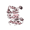
- PDB-5bpz: Atomic-resolution structures of the APC/C subunits Apc4 and the A... -
+ Open data

- Basic information
| Entry | Database: PDB / ID: 5bpz | ||||||
|---|---|---|---|---|---|---|---|
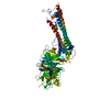
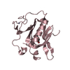
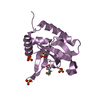
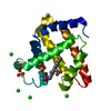
| Title | Atomic-resolution structures of the APC/C subunits Apc4 and the Apc5 N-terminal domain | ||||||
 Components Components | Anapc5 protein | ||||||
 Keywords Keywords | CELL CYCLE / Apc5 / APC/C / anaphase promoting complex | ||||||
| Function / homology |  Function and homology information Function and homology informationanaphase-promoting complex / anaphase-promoting complex-dependent catabolic process / positive regulation of mitotic metaphase/anaphase transition / protein K11-linked ubiquitination / spindle / cell division / cytoplasm Similarity search - Function | ||||||
| Biological species | |||||||
| Method |  X-RAY DIFFRACTION / SYNCHROTRON / Resolution: 2.18 Å X-RAY DIFFRACTION / SYNCHROTRON / Resolution: 2.18 Å | ||||||
 Authors Authors | Cronin, N. / Yang, J. / Zhang, Z. / Barford, D. | ||||||
| Funding support |  United Kingdom, 1items United Kingdom, 1items
| ||||||
 Citation Citation | Journal: J.Mol.Biol. / Year: 2015 Title: Atomic-Resolution Structures of the APC/C Subunits Apc4 and the Apc5 N-Terminal Domain. Authors: Cronin, N.B. / Yang, J. / Zhang, Z. / Kulkarni, K. / Chang, L. / Yamano, H. / Barford, D. | ||||||
| History |
|
- Structure visualization
| Structure viewer | Molecule: MolmilJmol/JSmol |
|---|
- Downloads & links
-Download
| PDBx/mmCIF format | 5bpz.cif.gz | 73.5 KB | Display | PDBx/mmCIF format |
|---|---|---|---|---|
| PDB format | pdb5bpz.ent.gz | 54.3 KB | Display | PDB format |
| PDBx/mmJSON format | 5bpz.json.gz | Tree view | PDBx/mmJSON format | |
| Others |  Other downloads Other downloads |
-Validation report
| Arichive directory | https://data.pdbj.org/pub/pdb/validation_reports/bp/5bpzftp://data.pdbj.org/pub/pdb/validation_reports/bp/5bpz | HTTPS FTP |
|---|
-Related structure data


-Links
 PDBj
PDBj- Assembly
| Deposited unit | 
| ||||||||
|---|---|---|---|---|---|---|---|---|---|
| 1 |
| ||||||||
| Unit cell |
|
-Components
| #1: Protein | Mass: 19453.600 Da / Num. of mol.: 1 / Fragment: N-terminus, UNP residues 1-162 Source method: isolated from a genetically manipulated source Source: (gene. exp.)  Trichoplusia ni (cabbage looper) / References: UniProt: Q0IH16 Trichoplusia ni (cabbage looper) / References: UniProt: Q0IH16 | ||
|---|---|---|---|
| #2: Chemical |   Mass: 62.068 Da / Num. of mol.: 2 / Source method: obtained synthetically / Formula: C2H6O2 Mass: 62.068 Da / Num. of mol.: 2 / Source method: obtained synthetically / Formula: C2H6O2#3: Water | ChemComp-HOH / |  Mass: 18.015 Da / Num. of mol.: 48 / Source method: isolated from a natural source / Formula: H2O Mass: 18.015 Da / Num. of mol.: 48 / Source method: isolated from a natural source / Formula: H2O |
-Experimental details
-Experiment
| Experiment | Method: X-RAY DIFFRACTION / Number of used crystals: 1 |
|---|
- Sample preparation
| Crystal | Density Matthews: 2.3 Å3/Da / Density % sol: 46.53 % / Description: Blocks |
|---|---|
| Crystal grow | Temperature: 293 K / Method: vapor diffusion, sitting drop / pH: 7 Details: The protein was concentrated to 3.4 mg/mL. Plate-like crystals were grown in a buffer containing 40 mM sodium propionate, 20 mM sodium cacodylate, 40 mM Bis-Tris propane (pH 7.0) and 25% ...Details: The protein was concentrated to 3.4 mg/mL. Plate-like crystals were grown in a buffer containing 40 mM sodium propionate, 20 mM sodium cacodylate, 40 mM Bis-Tris propane (pH 7.0) and 25% PEG1500. Larger crystals were obtained by seeding. |
-Data collection
| Diffraction | Mean temperature: 100 K |
|---|---|
| Diffraction source | Source: SYNCHROTRON / Site: Diamond / Beamline: I04 / Wavelength: 1.0081 Å |
| Detector | Type: ADSC QUANTUM 4 / Detector: CCD / Date: May 1, 2013 |
| Radiation | Protocol: SINGLE WAVELENGTH / Monochromatic (M) / Laue (L): M / Scattering type: x-ray |
| Radiation wavelength | Wavelength: 1.0081 Å / Relative weight: 1 |
| Reflection | Resolution: 2.18→63.63 Å / Num. all: 8213 / Num. obs: 8213 / % possible obs: 99.8 % / Observed criterion σ(F): 0 / Observed criterion σ(I): 0 / Redundancy: 12.8 % / Rmerge(I) obs: 0.055 / Net I/σ(I): 28.5 |
| Reflection shell | Resolution: 2.18→2.24 Å / Redundancy: 12.8 % / Rmerge(I) obs: 0.663 / Mean I/σ(I) obs: 4.5 / Num. unique all: 590 / % possible all: 99.1 |
- Processing
| Software |
| ||||||||||||||||||||||||||||||||||||||||||||||||||||||||||||||||||||||||||||||||||||||||||||||||||||
|---|---|---|---|---|---|---|---|---|---|---|---|---|---|---|---|---|---|---|---|---|---|---|---|---|---|---|---|---|---|---|---|---|---|---|---|---|---|---|---|---|---|---|---|---|---|---|---|---|---|---|---|---|---|---|---|---|---|---|---|---|---|---|---|---|---|---|---|---|---|---|---|---|---|---|---|---|---|---|---|---|---|---|---|---|---|---|---|---|---|---|---|---|---|---|---|---|---|---|---|---|---|
| Refinement | Resolution: 2.18→63 Å / Cor.coef. Fo:Fc: 0.9353 / Cor.coef. Fo:Fc free: 0.9208 / SU R Cruickshank DPI: 0.283 / Cross valid method: THROUGHOUT / σ(F): 0 / SU R Blow DPI: 0.303 / SU Rfree Blow DPI: 0.195 / SU Rfree Cruickshank DPI: 0.192
| ||||||||||||||||||||||||||||||||||||||||||||||||||||||||||||||||||||||||||||||||||||||||||||||||||||
| Displacement parameters | Biso max: 145.33 Å2 / Biso mean: 59.49 Å2 / Biso min: 27.89 Å2
| ||||||||||||||||||||||||||||||||||||||||||||||||||||||||||||||||||||||||||||||||||||||||||||||||||||
| Refine analyze | Luzzati coordinate error obs: 0.372 Å | ||||||||||||||||||||||||||||||||||||||||||||||||||||||||||||||||||||||||||||||||||||||||||||||||||||
| Refinement step | Cycle: final / Resolution: 2.18→63 Å
| ||||||||||||||||||||||||||||||||||||||||||||||||||||||||||||||||||||||||||||||||||||||||||||||||||||
| LS refinement shell | Resolution: 2.2→2.46 Å / Total num. of bins used: 5
| ||||||||||||||||||||||||||||||||||||||||||||||||||||||||||||||||||||||||||||||||||||||||||||||||||||
| Refinement TLS params. | Method: refined / Refine-ID: X-RAY DIFFRACTION
| ||||||||||||||||||||||||||||||||||||||||||||||||||||||||||||||||||||||||||||||||||||||||||||||||||||
| Refinement TLS group |
|
