[English] 日本語
Yorodumi
- PDB-5ll2: Structure of Isoleucine 2-epimerase from Lactobacillus buchneri (... -

+
Open data


ID or keywords:

Loading...

-
Basic information

Entry
Database: PDB / ID: 5ll2
TitleStructure of Isoleucine 2-epimerase from Lactobacillus buchneri (apo form)
ComponentsIsoleucine 2-epimerase
KeywordsISOMERASE / racemase / epimerase / Isoleucine / PLP / Lactobacillus buchneri
Function / homology
Function and homology information


isoleucine 2-epimerase / transaminase activity / isomerase activity / pyridoxal phosphate binding / identical protein binding
Similarity search - Function
: / Aminotransferases class-III pyridoxal-phosphate attachment site. / Aminotransferase class-III / Aminotransferase class-III / Aspartate Aminotransferase; domain 2 / Type I PLP-dependent aspartate aminotransferase-like (Major domain) / Pyridoxal phosphate-dependent transferase, small domain / Pyridoxal phosphate-dependent transferase, major domain / Pyridoxal phosphate-dependent transferase / 3-Layer(aba) Sandwich / Alpha Beta
Similarity search - Domain/homology
Isoleucine 2-epimerase
Similarity search - Component
Biological speciesLactobacillus buchneri (bacteria)
MethodX-RAY DIFFRACTION / SYNCHROTRON / MOLECULAR REPLACEMENT / Resolution: 2.6 Å
AuthorsReiser, J.-B. / Awad, R. / Gans, P.
Citation
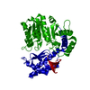
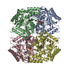
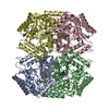
Journal: Biochimie / Year: 2017
Title: Structural insights into the substrate recognition and reaction specificity of the PLP-dependent fold-type I isoleucine 2-epimerase from Lactobacillus buchneri.
Authors: Awad, R. / Gans, P. / Reiser, J.B.
History
DepositionJul 26, 2016Deposition site: PDBE / Processing site: PDBE
Revision 1.0Apr 12, 2017Provider: repository / Type: Initial release
Revision 1.1May 17, 2017Group: Database references
Revision 1.2Aug 16, 2017Group: Data collection / Category: diffrn_source
Item: _diffrn_source.pdbx_synchrotron_beamline / _diffrn_source.type
Revision 1.3May 8, 2024Group: Data collection / Database references / Category: chem_comp_atom / chem_comp_bond / database_2
Item: _database_2.pdbx_DOI / _database_2.pdbx_database_accession

-
Structure visualization

Structure viewerMolecule:
MolmilJmol/JSmol

Downloads & links

-
Assembly

Deposited unit
A: Isoleucine 2-epimerase
B: Isoleucine 2-epimerase
C: Isoleucine 2-epimerase
D: Isoleucine 2-epimerase


Theoretical massNumber of molelcules
Total (without water)211,3874
Polymers211,3874
Non-polymers00
Water46826
1


  • Idetical with deposited unit
  • defined by author&software
TypeNameSymmetry operationNumber
identity operation1_555x,y,z1
Buried area17650 Å2
ΔGint-115 kcal/mol
Surface area54230 Å2
MethodPISA
Unit cell
Length a, b, c (Å)59.160, 161.780, 186.170
Angle α, β, γ (deg.)90.00, 90.00, 90.00
Int Tables number19
Space group name H-MP212121

-
Components

#1: Protein
Isoleucine 2-epimerase / BCAA racemase


Mass: 52846.770 Da / Num. of mol.: 4
Source method: isolated from a genetically manipulated source
Source: (gene. exp.) Lactobacillus buchneri (bacteria) / Production host: Escherichia coli BL21(DE3) (bacteria) / References: UniProt: M1GRN3, isoleucine 2-epimerase
#2: Water ChemComp-HOH / water


Mass: 18.015 Da / Num. of mol.: 26 / Source method: isolated from a natural source / Formula: H2O

-
Experimental details

-
Experiment

ExperimentMethod: X-RAY DIFFRACTION / Number of used crystals: 1

-
Sample preparation

CrystalDensity Matthews: 2.11 Å3/Da / Density % sol: 41.63 %
Crystal growTemperature: 293 K / Method: vapor diffusion, hanging drop / Details: 12 to 18 % PEG3350 and 100 mM Lithium Citrate

-
Data collection

DiffractionMean temperature: 100 K
Diffraction sourceSource: SYNCHROTRON / Site: ESRF / Beamline: MASSIF-3 / Wavelength: 0.97 Å
DetectorType: DECTRIS EIGER X 4M / Detector: PIXEL / Date: May 4, 2016
RadiationProtocol: SINGLE WAVELENGTH / Monochromatic (M) / Laue (L): M / Scattering type: x-ray
Radiation wavelengthWavelength: 0.97 Å / Relative weight: 1
ReflectionResolution: 2.6→20 Å / Num. obs: 55147 / % possible obs: 98.5 % / Redundancy: 3.9 % / Rmerge(I) obs: 0.12 / Rsym value: 0.12 / Net I/σ(I): 9.3
Reflection shellResolution: 2.6→2.7 Å / Redundancy: 4.2 % / Rmerge(I) obs: 0.616 / Mean I/σ(I) obs: 2 / % possible all: 98.1

-
Processing

Software
NameVersionClassification
REFMAC5.8.0135refinement
XDSdata reduction
XSCALEdata scaling
REFMACphasing
RefinementMethod to determine structure: MOLECULAR REPLACEMENT / Resolution: 2.6→19.93 Å / Cor.coef. Fo:Fc: 0.887 / Cor.coef. Fo:Fc free: 0.806 / SU B: 12.583 / SU ML: 0.286 / Cross valid method: THROUGHOUT / ESU R: 0.338 / ESU R Free: 0.087 / Stereochemistry target values: MAXIMUM LIKELIHOOD / Details: HYDROGENS HAVE BEEN USED IF PRESENT IN THE INPUT
RfactorNum. reflection% reflectionSelection details
Rfree0.31599 2752 5 %RANDOM
Rwork0.24387 ---
obs0.24747 52387 98.61 %-
Solvent computationIon probe radii: 0.8 Å / Shrinkage radii: 0.8 Å / VDW probe radii: 1.2 Å / Solvent model: BABINET MODEL WITH MASK
Displacement parametersBiso mean: 43.475 Å2
Baniso -1Baniso -2Baniso -3
1--0.05 Å20 Å20 Å2
2--0.04 Å20 Å2
3---0.01 Å2
Refinement stepCycle: LAST / Resolution: 2.6→19.93 Å
ProteinNucleic acidLigandSolventTotal
Num. atoms12549 0 0 26 12575
Refine LS restraints
Refine-IDTypeDev idealDev ideal targetNumber
X-RAY DIFFRACTIONr_bond_refined_d0.0130.01912838
X-RAY DIFFRACTIONr_bond_other_d
X-RAY DIFFRACTIONr_angle_refined_deg1.8421.96117407
X-RAY DIFFRACTIONr_angle_other_deg
X-RAY DIFFRACTIONr_dihedral_angle_1_deg8.22951624
X-RAY DIFFRACTIONr_dihedral_angle_2_deg37.90824.846551
X-RAY DIFFRACTIONr_dihedral_angle_3_deg21.424152142
X-RAY DIFFRACTIONr_dihedral_angle_4_deg17.4331544
X-RAY DIFFRACTIONr_chiral_restr0.1220.21933
X-RAY DIFFRACTIONr_gen_planes_refined0.0080.0219708
X-RAY DIFFRACTIONr_gen_planes_other
X-RAY DIFFRACTIONr_nbd_refined
X-RAY DIFFRACTIONr_nbd_other
X-RAY DIFFRACTIONr_nbtor_refined
X-RAY DIFFRACTIONr_nbtor_other
X-RAY DIFFRACTIONr_xyhbond_nbd_refined
X-RAY DIFFRACTIONr_xyhbond_nbd_other
X-RAY DIFFRACTIONr_metal_ion_refined
X-RAY DIFFRACTIONr_metal_ion_other
X-RAY DIFFRACTIONr_symmetry_vdw_refined
X-RAY DIFFRACTIONr_symmetry_vdw_other
X-RAY DIFFRACTIONr_symmetry_hbond_refined
X-RAY DIFFRACTIONr_symmetry_hbond_other
X-RAY DIFFRACTIONr_symmetry_metal_ion_refined
X-RAY DIFFRACTIONr_symmetry_metal_ion_other
X-RAY DIFFRACTIONr_mcbond_it2.9974.3016514
X-RAY DIFFRACTIONr_mcbond_other
X-RAY DIFFRACTIONr_mcangle_it4.7266.4428129
X-RAY DIFFRACTIONr_mcangle_other
X-RAY DIFFRACTIONr_scbond_it2.9344.3926324
X-RAY DIFFRACTIONr_scbond_other
X-RAY DIFFRACTIONr_scangle_it
X-RAY DIFFRACTIONr_scangle_other
X-RAY DIFFRACTIONr_long_range_B_refined8.63836.44819787
X-RAY DIFFRACTIONr_long_range_B_other
X-RAY DIFFRACTIONr_rigid_bond_restr
X-RAY DIFFRACTIONr_sphericity_free
X-RAY DIFFRACTIONr_sphericity_bonded
LS refinement shellResolution: 2.601→2.669 Å / Total num. of bins used: 20
RfactorNum. reflection% reflection
Rfree0.352 202 -
Rwork0.243 3817 -
obs--98.6 %

+
About Yorodumi

-
News

-
Feb 9, 2022. New format data for meta-information of EMDB entries

New format data for meta-information of EMDB entries

  • Version 3 of the EMDB header file is now the official format.
  • The previous official version 1.9 will be removed from the archive.

Related info.:EMDB header

External links:wwPDB to switch to version 3 of the EMDB data model

-
Aug 12, 2020. Covid-19 info

Covid-19 info

URL: https://pdbjlvh1.pdbj.org/emnavi/covid19.php

New page: Covid-19 featured information page in EM Navigator.

Related info.:Covid-19 info / Mar 5, 2020. Novel coronavirus structure data

+
Mar 5, 2020. Novel coronavirus structure data

Novel coronavirus structure data

Related info.:Yorodumi Speices / Aug 12, 2020. Covid-19 info

External links:COVID-19 featured content - PDBj / Molecule of the Month (242):Coronavirus Proteases

+
Jan 31, 2019. EMDB accession codes are about to change! (news from PDBe EMDB page)

EMDB accession codes are about to change! (news from PDBe EMDB page)

  • The allocation of 4 digits for EMDB accession codes will soon come to an end. Whilst these codes will remain in use, new EMDB accession codes will include an additional digit and will expand incrementally as the available range of codes is exhausted. The current 4-digit format prefixed with “EMD-” (i.e. EMD-XXXX) will advance to a 5-digit format (i.e. EMD-XXXXX), and so on. It is currently estimated that the 4-digit codes will be depleted around Spring 2019, at which point the 5-digit format will come into force.
  • The EM Navigator/Yorodumi systems omit the EMD- prefix.

Related info.:Q: What is EMD? / ID/Accession-code notation in Yorodumi/EM Navigator

External links:EMDB Accession Codes are Changing Soon! / Contact to PDBj

+
Jul 12, 2017. Major update of PDB

Major update of PDB

  • wwPDB released updated PDB data conforming to the new PDBx/mmCIF dictionary.
  • This is a major update changing the version number from 4 to 5, and with Remediation, in which all the entries are updated.
  • In this update, many items about electron microscopy experimental information are reorganized (e.g. em_software).
  • Now, EM Navigator and Yorodumi are based on the updated data.

External links:wwPDB Remediation / Enriched Model Files Conforming to OneDep Data Standards Now Available in the PDB FTP Archive

-
Yorodumi

Thousand views of thousand structures

  • Yorodumi is a browser for structure data from EMDB, PDB, SASBDB, etc.
  • This page is also the successor to EM Navigator detail page, and also detail information page/front-end page for Omokage search.
  • The word "yorodu" (or yorozu) is an old Japanese word meaning "ten thousand". "mi" (miru) is to see.

Related info.:EMDB / PDB / SASBDB / Comparison of 3 databanks / Yorodumi Search / Aug 31, 2016. New EM Navigator & Yorodumi / Yorodumi Papers / Jmol/JSmol / Function and homology information / Changes in new EM Navigator and Yorodumi

Read more