 Movie
Movie Controller

[English] 日本語
 Yorodumi
- PDB-5lhv: X-ray structure of uridine phosphorylase from Vibrio cholerae in ... -
+ Open data

- Basic information
| Entry | Database: PDB / ID: 5lhv | ||||||
|---|---|---|---|---|---|---|---|
| Title | X-ray structure of uridine phosphorylase from Vibrio cholerae in complex with uridine and sulfate ion at 1.29 A resolution | ||||||
 Components Components | Uridine phosphorylase | ||||||
 Keywords Keywords | TRANSFERASE / Rossmann Fold | ||||||
| Function / homology |  Function and homology information Function and homology informationnucleotide catabolic process / uridine phosphorylase / nucleoside catabolic process / UMP salvage / uridine phosphorylase activity / metal ion binding / cytosol Similarity search - Function | ||||||
| Biological species |   Vibrio cholerae (bacteria) Vibrio cholerae (bacteria) | ||||||
| Method |  X-RAY DIFFRACTION / SYNCHROTRON / MOLECULAR REPLACEMENT / molecular replacement / Resolution: 1.288 Å X-RAY DIFFRACTION / SYNCHROTRON / MOLECULAR REPLACEMENT / molecular replacement / Resolution: 1.288 Å | ||||||
 Authors Authors | Prokofev, I.I. / Lashkov, A.A. / Gabdoulkhakov, A.G. / Balaev, V.V. / Betzel, C. / Mikhailov, A.M. | ||||||
| Funding support |  Russian Federation, 1items Russian Federation, 1items
| ||||||
 Citation Citation | Journal: To Be Published Title: X-ray structure of uridine phosphorylase from Vibrio cholerae in complex with uridine and sulfate ion at 1.29 A resolution Authors: Prokofev, I.I. / Lashkov, A.A. / Gabdoulkhakov, A.G. / Balaev, V.V. / Betzel, C. / Mikhailov, A.M. | ||||||
| History |
|
- Structure visualization
| Structure viewer | Molecule: MolmilJmol/JSmol |
|---|
- Downloads & links
-Download
| PDBx/mmCIF format | 5lhv.cif.gz | 667.4 KB | Display | PDBx/mmCIF format |
|---|---|---|---|---|
| PDB format | pdb5lhv.ent.gz | 547.6 KB | Display | PDB format |
| PDBx/mmJSON format | 5lhv.json.gz | Tree view | PDBx/mmJSON format | |
| Others |  Other downloads Other downloads |
-Validation report
| Arichive directory | https://data.pdbj.org/pub/pdb/validation_reports/lh/5lhvftp://data.pdbj.org/pub/pdb/validation_reports/lh/5lhv | HTTPS FTP |
|---|
-Related structure data
| Related structure data |  4k6oS S: Starting model for refinement |
|---|---|
| Similar structure data |


-Links
 PDBj
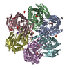
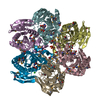
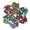
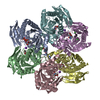
PDBj- Assembly
| Deposited unit | 
| ||||||||
|---|---|---|---|---|---|---|---|---|---|
| 1 |
| ||||||||
| Unit cell |
|
-Components
-Protein , 1 types, 6 molecules ABCDEF
| #1: Protein | Mass: 26876.693 Da / Num. of mol.: 6 Source method: isolated from a genetically manipulated source Source: (gene. exp.) Vibrio cholerae (bacteria)Gene: udp, udp_1, DN30_1909, EN12_05055, ERS013138_02408, ERS013140_00580, ERS013186_00327, ERS013199_00063, ERS013201_00032, ERS013202_00369, ERS013206_00377 Production host: |
|---|
-Non-polymers , 8 types, 1629 molecules 


| #2: Chemical | ChemComp-SO4 /  Mass: 96.063 Da / Num. of mol.: 8 / Source method: obtained synthetically / Formula: SO4 Mass: 96.063 Da / Num. of mol.: 8 / Source method: obtained synthetically / Formula: SO4#3: Chemical | ChemComp-URI /  Mass: 244.201 Da / Num. of mol.: 6 / Source method: obtained synthetically / Formula: C9H12N2O6 Mass: 244.201 Da / Num. of mol.: 6 / Source method: obtained synthetically / Formula: C9H12N2O6#4: Chemical | ChemComp-URA /  Mass: 112.087 Da / Num. of mol.: 6 / Source method: obtained synthetically / Formula: C4H4N2O2 Mass: 112.087 Da / Num. of mol.: 6 / Source method: obtained synthetically / Formula: C4H4N2O2#5: Chemical | ChemComp-MG /  Mass: 24.305 Da / Num. of mol.: 7 / Source method: obtained synthetically / Formula: Mg Mass: 24.305 Da / Num. of mol.: 7 / Source method: obtained synthetically / Formula: Mg#6: Chemical | ChemComp-CL /  Mass: 35.453 Da / Num. of mol.: 6 / Source method: obtained synthetically / Formula: Cl Mass: 35.453 Da / Num. of mol.: 6 / Source method: obtained synthetically / Formula: Cl#7: Chemical |  Mass: 22.990 Da / Num. of mol.: 3 / Source method: obtained synthetically / Formula: Na Mass: 22.990 Da / Num. of mol.: 3 / Source method: obtained synthetically / Formula: Na#8: Chemical | ChemComp-GOL /  Mass: 92.094 Da / Num. of mol.: 7 / Source method: obtained synthetically / Formula: C3H8O3 Mass: 92.094 Da / Num. of mol.: 7 / Source method: obtained synthetically / Formula: C3H8O3#9: Water | ChemComp-HOH / | Mass: 18.015 Da / Num. of mol.: 1586 / Source method: isolated from a natural source / Formula: H2O |
|---|
-Experimental details
-Experiment
| Experiment | Method: X-RAY DIFFRACTION / Number of used crystals: 1 |
|---|
- Sample preparation
| Crystal | Density Matthews: 2.25 Å3/Da / Density % sol: 45.32 % |
|---|---|
| Crystal grow | Temperature: 293 K / Method: vapor diffusion, hanging drop / pH: 8.5 / Details: PEG4000, 0.1M TRIS-HCl, 0.2M MgCl2x6H2O |
-Data collection
| Diffraction | Mean temperature: 100 K | ||||||||||||||||||||||||||||||||||||||||||||||||||||||||||||||||||||||
|---|---|---|---|---|---|---|---|---|---|---|---|---|---|---|---|---|---|---|---|---|---|---|---|---|---|---|---|---|---|---|---|---|---|---|---|---|---|---|---|---|---|---|---|---|---|---|---|---|---|---|---|---|---|---|---|---|---|---|---|---|---|---|---|---|---|---|---|---|---|---|---|
| Diffraction source | Source: SYNCHROTRON / Site: PETRA III, DESY  / Beamline: P11 / Wavelength: 1 Å / Beamline: P11 / Wavelength: 1 Å | ||||||||||||||||||||||||||||||||||||||||||||||||||||||||||||||||||||||
| Detector | Type: PSI PILATUS 6M / Detector: PIXEL / Date: Apr 17, 2016 | ||||||||||||||||||||||||||||||||||||||||||||||||||||||||||||||||||||||
| Radiation | Protocol: SINGLE WAVELENGTH / Monochromatic (M) / Laue (L): M / Scattering type: x-ray | ||||||||||||||||||||||||||||||||||||||||||||||||||||||||||||||||||||||
| Radiation wavelength | Wavelength: 1 Å / Relative weight: 1 | ||||||||||||||||||||||||||||||||||||||||||||||||||||||||||||||||||||||
| Reflection | Resolution: 1.288→46.23 Å / Num. obs: 337058 / % possible obs: 94 % / Observed criterion σ(I): -3 / Redundancy: 3.5 % / Biso Wilson estimate: 10.77 Å2 / CC1/2: 0.999 / Rmerge(I) obs: 0.059 / Net I/σ(I): 13.18 | ||||||||||||||||||||||||||||||||||||||||||||||||||||||||||||||||||||||
| Reflection shell |
|
-Phasing
| Phasing | Method: molecular replacement | ||||||
|---|---|---|---|---|---|---|---|
| Phasing MR | R rigid body: 0.525
|
- Processing
| Software |
| ||||||||||||||||||||||||||||||||||||||||||||||||||||||||||||||||||||||||||||||||||||||||||||||||||||||||||||||||||||||||||||||||||||||||||||||||||||||||||||||||||||||||||||||||||||||
|---|---|---|---|---|---|---|---|---|---|---|---|---|---|---|---|---|---|---|---|---|---|---|---|---|---|---|---|---|---|---|---|---|---|---|---|---|---|---|---|---|---|---|---|---|---|---|---|---|---|---|---|---|---|---|---|---|---|---|---|---|---|---|---|---|---|---|---|---|---|---|---|---|---|---|---|---|---|---|---|---|---|---|---|---|---|---|---|---|---|---|---|---|---|---|---|---|---|---|---|---|---|---|---|---|---|---|---|---|---|---|---|---|---|---|---|---|---|---|---|---|---|---|---|---|---|---|---|---|---|---|---|---|---|---|---|---|---|---|---|---|---|---|---|---|---|---|---|---|---|---|---|---|---|---|---|---|---|---|---|---|---|---|---|---|---|---|---|---|---|---|---|---|---|---|---|---|---|---|---|---|---|---|---|
| Refinement | Method to determine structure: MOLECULAR REPLACEMENT Starting model: 4K6O Resolution: 1.288→33.569 Å / SU ML: 0.14 / Cross valid method: THROUGHOUT / σ(F): 1.95 / Phase error: 20.29
| ||||||||||||||||||||||||||||||||||||||||||||||||||||||||||||||||||||||||||||||||||||||||||||||||||||||||||||||||||||||||||||||||||||||||||||||||||||||||||||||||||||||||||||||||||||||
| Solvent computation | Shrinkage radii: 0.9 Å / VDW probe radii: 1.11 Å | ||||||||||||||||||||||||||||||||||||||||||||||||||||||||||||||||||||||||||||||||||||||||||||||||||||||||||||||||||||||||||||||||||||||||||||||||||||||||||||||||||||||||||||||||||||||
| Displacement parameters | Biso max: 70.01 Å2 / Biso mean: 22.24 Å2 / Biso min: 7.15 Å2 | ||||||||||||||||||||||||||||||||||||||||||||||||||||||||||||||||||||||||||||||||||||||||||||||||||||||||||||||||||||||||||||||||||||||||||||||||||||||||||||||||||||||||||||||||||||||
| Refinement step | Cycle: final / Resolution: 1.288→33.569 Å
| ||||||||||||||||||||||||||||||||||||||||||||||||||||||||||||||||||||||||||||||||||||||||||||||||||||||||||||||||||||||||||||||||||||||||||||||||||||||||||||||||||||||||||||||||||||||
| Refine LS restraints |
| ||||||||||||||||||||||||||||||||||||||||||||||||||||||||||||||||||||||||||||||||||||||||||||||||||||||||||||||||||||||||||||||||||||||||||||||||||||||||||||||||||||||||||||||||||||||
| LS refinement shell | Refine-ID: X-RAY DIFFRACTION / Total num. of bins used: 25
|
