 Movie
Movie Controller

[English] 日本語
 Yorodumi
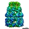
- PDB-5ldf: Maltose binding protein genetically fused to dodecameric glutamin... -
+ Open data

- Basic information
| Entry | Database: PDB / ID: 5ldf | |||||||||
|---|---|---|---|---|---|---|---|---|---|---|
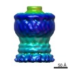
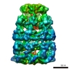
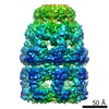
| Title | Maltose binding protein genetically fused to dodecameric glutamine synthetase | |||||||||
 Components Components |
| |||||||||
 Keywords Keywords | LIGASE / Fusion protein / chimera / dodecamer / symmetrized construct | |||||||||
| Function / homology |  Function and homology information Function and homology informationnitrogen utilization / glutamine synthetase / : / glutamine synthetase activity / carbohydrate transmembrane transporter activity / maltose binding / maltose transport / maltodextrin transmembrane transport / ATP-binding cassette (ABC) transporter complex, substrate-binding subunit-containing / outer membrane-bounded periplasmic space ...nitrogen utilization / glutamine synthetase / : / glutamine synthetase activity / carbohydrate transmembrane transporter activity / maltose binding / maltose transport / maltodextrin transmembrane transport / ATP-binding cassette (ABC) transporter complex, substrate-binding subunit-containing / outer membrane-bounded periplasmic space / ATP binding / membrane / metal ion binding / cytoplasm Similarity search - Function | |||||||||
| Biological species |  Salmonella typhi (bacteria) Salmonella typhi (bacteria) | |||||||||
| Method | ELECTRON MICROSCOPY / single particle reconstruction / cryo EM / Resolution: 6.2 Å | |||||||||
 Authors Authors | Coscia, F. / Petosa, C. / Schoehn, G. | |||||||||
| Funding support |  France, 1items France, 1items
| |||||||||
 Citation Citation | Journal: Sci Rep / Year: 2016 Title: Fusion to a homo-oligomeric scaffold allows cryo-EM analysis of a small protein. Authors: Francesca Coscia / Leandro F Estrozi / Fabienne Hans / Hélène Malet / Marjolaine Noirclerc-Savoye / Guy Schoehn / Carlo Petosa / Abstract: Recent technical advances have revolutionized the field of cryo-electron microscopy (cryo-EM). However, most monomeric proteins remain too small (<100 kDa) for cryo-EM analysis. To overcome this limitation, we explored a strategy whereby a monomeric target protein is genetically fused to a homo-oligomeric scaffold protein and the junction optimized to allow the target to adopt the scaffold symmetry, thereby generating a chimeric particle suitable for cryo-EM. To demonstrate the concept, we fused maltose-binding protein (MBP), a 40 kDa monomer, to glutamine synthetase, a dodecamer formed by two hexameric rings. Chimeric constructs with different junction lengths were screened by biophysical analysis and negative-stain EM. The optimal construct yielded a cryo-EM reconstruction that revealed the MBP structure at sub-nanometre resolution. These findings illustrate the feasibility of using homo-oligomeric scaffolds to enable cryo-EM analysis of monomeric proteins, paving the way for applying this strategy to challenging structures resistant to crystallographic and NMR analysis. | |||||||||
| History |
|
- Structure visualization
| Movie |
Movie viewer |
|---|---|
| Structure viewer | Molecule: MolmilJmol/JSmol |
- Downloads & links
-Download
| PDBx/mmCIF format | 5ldf.cif.gz | 1.6 MB | Display | PDBx/mmCIF format |
|---|---|---|---|---|
| PDB format | pdb5ldf.ent.gz | 1.3 MB | Display | PDB format |
| PDBx/mmJSON format | 5ldf.json.gz | Tree view | PDBx/mmJSON format | |
| Others |  Other downloads Other downloads |
-Validation report
| Arichive directory | https://data.pdbj.org/pub/pdb/validation_reports/ld/5ldfftp://data.pdbj.org/pub/pdb/validation_reports/ld/5ldf | HTTPS FTP |
|---|
-Related structure data
| Related structure data | 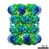 4039MC M: map data used to model this data C: citing same article ( |
|---|---|
| Similar structure data |
-Links
 PDBj
PDBj


- Assembly
| Deposited unit | 
|
|---|---|
| 1 |
|
-Components
| #1: Protein | Mass: 51586.266 Da / Num. of mol.: 12 / Mutation: Deletion of residues 1-2 Source method: isolated from a genetically manipulated source Source: (gene. exp.) Salmonella typhi (bacteria) / Gene: glnA, STY3874, t3614 / Production host: #2: Protein | Mass: 40753.152 Da / Num. of mol.: 12 Source method: isolated from a genetically manipulated source Source: (gene. exp.) #3: Polysaccharide | alpha-D-glucopyranose-(1-4)-alpha-D-glucopyranose / alpha-maltose   Source method: isolated from a genetically manipulated source Details: oligosaccharide / References: alpha-maltose |
|---|
-Experimental details
-Experiment
| Experiment | Method: ELECTRON MICROSCOPY |
|---|---|
| EM experiment | Aggregation state: PARTICLE / 3D reconstruction method: single particle reconstruction |
- Sample preparation
| Component | Name: Maltose-binding protein genetically fused to glutamine synthetase Type: COMPLEX / Entity ID: #1-#2 / Source: RECOMBINANT | |||||||||||||||||||||||||
|---|---|---|---|---|---|---|---|---|---|---|---|---|---|---|---|---|---|---|---|---|---|---|---|---|---|---|
| Molecular weight | Value: 1.11 MDa / Experimental value: NO | |||||||||||||||||||||||||
| Source (natural) | Organism: | |||||||||||||||||||||||||
| Source (recombinant) | Organism: | |||||||||||||||||||||||||
| Buffer solution | pH: 8 | |||||||||||||||||||||||||
| Buffer component |
| |||||||||||||||||||||||||
| Specimen | Conc.: 0.5 mg/ml / Embedding applied: NO / Shadowing applied: NO / Staining applied: NO / Vitrification applied: YES | |||||||||||||||||||||||||
| Specimen support | Grid material: COPPER/RHODIUM / Grid mesh size: 400 divisions/in. / Grid type: Quantifoil | |||||||||||||||||||||||||
| Vitrification | Instrument: FEI VITROBOT MARK IV / Cryogen name: ETHANE / Humidity: 100 % / Chamber temperature: 290 K |
- Electron microscopy imaging
| Experimental equipment |  Model: Tecnai F30 / Image courtesy: FEI Company |
|---|---|
| Microscopy | Model: FEI TECNAI F30 / Details: Polara Top entry |
| Electron gun | Electron source:  FIELD EMISSION GUN / Accelerating voltage: 300 kV / Illumination mode: FLOOD BEAM FIELD EMISSION GUN / Accelerating voltage: 300 kV / Illumination mode: FLOOD BEAM |
| Electron lens | Mode: BRIGHT FIELD / Nominal defocus max: 3500 nm / Nominal defocus min: 1000 nm / Alignment procedure: COMA FREE |
| Specimen holder | Cryogen: NITROGEN |
| Image recording | Average exposure time: 6 sec. / Electron dose: 25 e/Å2 / Detector mode: SUPER-RESOLUTION / Film or detector model: GATAN K2 SUMMIT (4k x 4k) / Num. of real images: 165 |
| Image scans | Movie frames/image: 40 |
- Processing
| EM software |
| ||||||||||||||||||||||||||||||||||||
|---|---|---|---|---|---|---|---|---|---|---|---|---|---|---|---|---|---|---|---|---|---|---|---|---|---|---|---|---|---|---|---|---|---|---|---|---|---|
| CTF correction | Type: PHASE FLIPPING ONLY | ||||||||||||||||||||||||||||||||||||
| Particle selection | Num. of particles selected: 39167 | ||||||||||||||||||||||||||||||||||||
| Symmetry | Point symmetry: D6 (2x6 fold dihedral) | ||||||||||||||||||||||||||||||||||||
| 3D reconstruction | Resolution: 6.2 Å / Resolution method: FSC 0.143 CUT-OFF / Num. of particles: 13847 / Symmetry type: POINT | ||||||||||||||||||||||||||||||||||||
| Atomic model building | Protocol: RIGID BODY FIT / Space: REAL |
