 Movie
Movie Controller

[English] 日本語
 Yorodumi
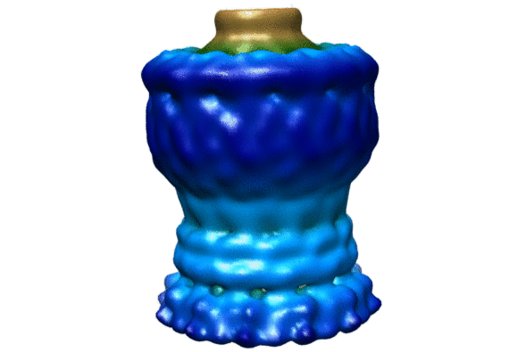
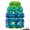
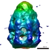
- EMDB-1763: 3D reconstruction of the type ii secretin GspD from Vibrio cholera -
+ Open data

- Basic information
| Entry | Database: EMDB / ID: EMD-1763 | |||||||||
|---|---|---|---|---|---|---|---|---|---|---|
| Title | 3D reconstruction of the type ii secretin GspD from Vibrio cholera | |||||||||
 Map data Map data | cryoEM 3D reconstruction of the type ii secretin GspD from vibrio cholera | |||||||||
 Sample Sample |
| |||||||||
 Keywords Keywords | Secretin / GspD / EpsD / type 2 secretion system / T2SS / outermembrane channel | |||||||||
| Function / homology | Type II secretion system protein GspD, conserved site / protein secretion Function and homology information Function and homology information | |||||||||
| Biological species |   Vibrio cholerae (bacteria) Vibrio cholerae (bacteria) | |||||||||
| Method | single particle reconstruction / cryo EM / Resolution: 19.0 Å | |||||||||
 Authors Authors | Reichow SL / Korotkov KV / Hol WGJ / Gonen T | |||||||||
 Citation Citation | Journal: Nat Struct Mol Biol / Year: 2010 Title: Structure of the cholera toxin secretion channel in its closed state. Authors: Steve L Reichow / Konstantin V Korotkov / Wim G J Hol / Tamir Gonen /  Abstract: The type II secretion system (T2SS) is a macromolecular complex spanning the inner and outer membranes of Gram-negative bacteria. Remarkably, the T2SS secretes folded proteins, including multimeric ...The type II secretion system (T2SS) is a macromolecular complex spanning the inner and outer membranes of Gram-negative bacteria. Remarkably, the T2SS secretes folded proteins, including multimeric assemblies such as cholera toxin and heat-labile enterotoxin from Vibrio cholerae and enterotoxigenic Escherichia coli, respectively. The major outer membrane T2SS protein is the 'secretin' GspD. Cryo-EM reconstruction of the V. cholerae secretin at 19-Å resolution reveals a dodecameric structure reminiscent of a barrel, with a large channel at its center that contains a closed periplasmic gate. The GspD periplasmic domain forms a vestibule with a conserved constriction, and it binds to a pentameric exoprotein and to the trimeric tip of the T2SS pseudopilus. By combining our results with structures of the cholera toxin and T2SS pseudopilus tip, we provide a structural basis for a possible secretion mechanism of the T2SS. | |||||||||
| History |
|
- Structure visualization
| Movie |
Movie viewer |
|---|---|
| Structure viewer | EM map: SurfViewMolmilJmol/JSmol |
| Supplemental images |

- Downloads & links
-EMDB archive
| Map data | emd_1763.map.gz | 10.4 MB | EMDB map data format | |
|---|---|---|---|---|
| Header (meta data) | emd-1763-v30.xmlemd-1763.xml | 9 KB 9 KB | Display Display | EMDB header |
| Images |  1763.gif 1763.gif EMD-1763.jpg EMD-1763.jpg | 52.9 KB 35.5 KB | ||
| Archive directory |  http://ftp.pdbj.org/pub/emdb/structures/EMD-1763ftp://ftp.pdbj.org/pub/emdb/structures/EMD-1763 http://ftp.pdbj.org/pub/emdb/structures/EMD-1763ftp://ftp.pdbj.org/pub/emdb/structures/EMD-1763 | HTTPS FTP |
-Related structure data
| Similar structure data |
|---|
-Links
| EMDB pages | EMDB (EBI/PDBe) / EMDataResource |
|---|
-Map
| File | Download / File: emd_1763.map.gz / Format: CCP4 / Size: 12.6 MB / Type: IMAGE STORED AS FLOATING POINT NUMBER (4 BYTES) | ||||||||||||||||||||||||||||||||||||||||||||||||||||||||||||||||||||
|---|---|---|---|---|---|---|---|---|---|---|---|---|---|---|---|---|---|---|---|---|---|---|---|---|---|---|---|---|---|---|---|---|---|---|---|---|---|---|---|---|---|---|---|---|---|---|---|---|---|---|---|---|---|---|---|---|---|---|---|---|---|---|---|---|---|---|---|---|---|
| Annotation | cryoEM 3D reconstruction of the type ii secretin GspD from vibrio cholera | ||||||||||||||||||||||||||||||||||||||||||||||||||||||||||||||||||||
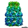
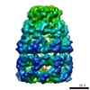
| Projections & slices | Image control
Images are generated by Spider. | ||||||||||||||||||||||||||||||||||||||||||||||||||||||||||||||||||||
| Voxel size | X=Y=Z: 2.54 Å | ||||||||||||||||||||||||||||||||||||||||||||||||||||||||||||||||||||
| Density |
| ||||||||||||||||||||||||||||||||||||||||||||||||||||||||||||||||||||
| Symmetry | Space group: 1 | ||||||||||||||||||||||||||||||||||||||||||||||||||||||||||||||||||||
| Details | EMDB XML:
CCP4 map header:
| ||||||||||||||||||||||||||||||||||||||||||||||||||||||||||||||||||||
 Z (Sec.)
 Y (Row.)
 X (Col.)


-Supplemental data
- Sample components
-Entire : Type ii secretin GspD from Vibrio cholera
| Entire | Name: Type ii secretin GspD from Vibrio cholera |
|---|---|
| Components |
|
-Supramolecule #1000: Type ii secretin GspD from Vibrio cholera
| Supramolecule | Name: Type ii secretin GspD from Vibrio cholera / type: sample / ID: 1000 / Oligomeric state: Dodecamer / Number unique components: 1 |
|---|---|
| Molecular weight | Experimental: 900 KDa / Theoretical: 900 KDa / Method: Size-exclusion chromatography |
-Macromolecule #1: General Secretion Protein D
| Macromolecule | Name: General Secretion Protein D / type: protein_or_peptide / ID: 1 / Name.synonym: GspD / Number of copies: 12 / Oligomeric state: Dodecamer / Recombinant expression: Yes |
|---|---|
| Source (natural) | Organism: Vibrio cholerae (bacteria) / Location in cell: Membrane |
| Molecular weight | Experimental: 900 KDa / Theoretical: 900 KDa |
| Recombinant expression | Organism: |
| Sequence | GO: protein secretion InterPro: Type II secretion system protein GspD, conserved site |
-Experimental details
-Structure determination
| Method | cryo EM |
|---|---|
 Processing Processing | single particle reconstruction |
| Aggregation state | particle |
-Sample preparation
| Concentration | 0.05 mg/mL |
|---|---|
| Buffer | pH: 7.8 Details: 20mM Tris-HCl pH 7.8, 300mM NaCl, N-dodecyl-N,N-dimethylammonio-1-propanesulfonate (SB3-12) |
| Grid | Details: Holey carbon grid with cont. carbon overlayed |
| Vitrification | Cryogen name: ETHANE / Chamber humidity: 100 % / Instrument: OTHER / Details: Vitrification instrument: Vitrobot / Method: Blot with filter paper and plunge to liquid ethane |
- Electron microscopy
| Microscope | FEI TECNAI F20 |
|---|---|
| Image recording | Category: FILM / Film or detector model: KODAK SO-163 FILM / Digitization - Scanner: NIKON SUPER COOLSCAN 9000 / Digitization - Sampling interval: 6.9 µm Details: Images were binned 2 times after scanning to yield a final pixel size of 2.54 angstrom |
| Tilt angle min | 0 |
| Tilt angle max | 0 |
| Electron beam | Acceleration voltage: 200 kV / Electron source:  FIELD EMISSION GUN FIELD EMISSION GUN |
| Electron optics | Calibrated magnification: 51159 / Illumination mode: SPOT SCAN / Imaging mode: BRIGHT FIELD / Nominal defocus max: 5.0 µm / Nominal defocus min: 1.5 µm / Nominal magnification: 50000 |
| Sample stage | Specimen holder: Gatan / Specimen holder model: GATAN LIQUID NITROGEN |
| Experimental equipment |  Model: Tecnai F20 / Image courtesy: FEI Company |
-Image processing
| Details | Particles were selected by hand |
|---|---|
| CTF correction | Details: CTFTILT |
| Final reconstruction | Applied symmetry - Point group: C12 (12 fold cyclic) / Resolution.type: BY AUTHOR / Resolution: 19.0 Å / Resolution method: FSC 0.5 CUT-OFF / Software - Name: FREALIGN / Number images used: 10000 |
