[English] 日本語
Yorodumi
- PDB-5ld5: Crystal structure of a bacterial dehydrogenase at 2.19 Angstroms ... -

+
Open data


ID or keywords:

Loading...

-
Basic information

Entry
Database: PDB / ID: 5ld5
Title
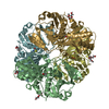
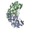
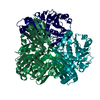
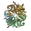
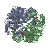
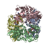
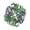
Crystal structure of a bacterial dehydrogenase at 2.19 Angstroms resolution
ComponentsGlyceraldehyde-3-phosphate dehydrogenaseGlyceraldehyde 3-phosphate dehydrogenase
KeywordsOXIDOREDUCTASE / Atopobium vaginae / GAPDH / NAD / dehydrogenase
Function / homology
Function and homology information


Oxidoreductases; Acting on the aldehyde or oxo group of donors; With NAD+ or NADP+ as acceptor / oxidoreductase activity, acting on the aldehyde or oxo group of donors, NAD or NADP as acceptor / glucose metabolic process / NAD binding / NADP binding
Similarity search - Function
Glyceraldehyde-3-phosphate dehydrogenase, type I / Glyceraldehyde 3-phosphate dehydrogenase, active site / Glyceraldehyde 3-phosphate dehydrogenase active site. / Glyceraldehyde 3-phosphate dehydrogenase, NAD binding domain / Glyceraldehyde 3-phosphate dehydrogenase, NAD(P) binding domain / Glyceraldehyde 3-phosphate dehydrogenase, catalytic domain / Glyceraldehyde/Erythrose phosphate dehydrogenase family / Glyceraldehyde 3-phosphate dehydrogenase, C-terminal domain / Glyceraldehyde 3-phosphate dehydrogenase, NAD binding domain / Dihydrodipicolinate Reductase; domain 2 ...Glyceraldehyde-3-phosphate dehydrogenase, type I / Glyceraldehyde 3-phosphate dehydrogenase, active site / Glyceraldehyde 3-phosphate dehydrogenase active site. / Glyceraldehyde 3-phosphate dehydrogenase, NAD binding domain / Glyceraldehyde 3-phosphate dehydrogenase, NAD(P) binding domain / Glyceraldehyde 3-phosphate dehydrogenase, catalytic domain / Glyceraldehyde/Erythrose phosphate dehydrogenase family / Glyceraldehyde 3-phosphate dehydrogenase, C-terminal domain / Glyceraldehyde 3-phosphate dehydrogenase, NAD binding domain / Dihydrodipicolinate Reductase; domain 2 / Dihydrodipicolinate Reductase; domain 2 / NAD(P)-binding domain superfamily / 2-Layer Sandwich / Alpha Beta
Similarity search - Domain/homology
NICOTINAMIDE-ADENINE-DINUCLEOTIDE / Glyceraldehyde-3-phosphate dehydrogenase
Similarity search - Component
Biological speciesAtopobium vaginae (bacteria)
MethodX-RAY DIFFRACTION / SYNCHROTRON / SAD / Resolution: 2.1906 Å
AuthorsQuerol-Garcia, J. / Fernandez, F.J. / Gomez, S. / Fulla, D. / Juanhuix, J. / Vega, M.C.
Funding support Spain, Belgium, 4items
OrganizationGrant numberCountry
Spanish Ministry of Economy and CompetitivenessCTQ2015-66206-C2-2-R Spain
Instituto de Salud Carlos IIIPI12/01667 Spain
Regional Government of MadridS2010/BD-2316 Spain
European UnionComplexINC (Contract No. 279039) Belgium
CitationJournal: Front Microbiol / Year: 2017
Title: Crystal Structure of Glyceraldehyde-3-Phosphate Dehydrogenase from the Gram-Positive Bacterial Pathogen A. vaginae, an Immunoevasive Factor that Interacts with the Human C5a Anaphylatoxin.
Authors: Querol-Garcia, J. / Fernandez, F.J. / Marin, A.V. / Gomez, S. / Fulla, D. / Melchor-Tafur, C. / Franco-Hidalgo, V. / Alberti, S. / Juanhuix, J. / Rodriguez de Cordoba, S. / Regueiro, J.R. / Vega, M.C.
History
DepositionJun 23, 2016Deposition site: PDBE / Processing site: PDBE
Revision 1.0Jul 12, 2017Provider: repository / Type: Initial release
Revision 1.1Sep 13, 2017Group: Author supporting evidence / Category: pdbx_audit_support / Item: _pdbx_audit_support.funding_organization
Revision 1.2May 8, 2024Group: Data collection / Database references / Category: chem_comp_atom / chem_comp_bond / database_2
Item: _database_2.pdbx_DOI / _database_2.pdbx_database_accession

-
Structure visualization

Structure viewerMolecule:
MolmilJmol/JSmol

Downloads & links

-
Assembly

Deposited unit
A: Glyceraldehyde-3-phosphate dehydrogenase
B: Glyceraldehyde-3-phosphate dehydrogenase
C: Glyceraldehyde-3-phosphate dehydrogenase
D: Glyceraldehyde-3-phosphate dehydrogenase
hetero molecules


Theoretical massNumber of molelcules
Total (without water)157,60520
Polymers153,8464
Non-polymers3,75916
Water5,855325
1


  • Idetical with deposited unit
  • defined by author&software
TypeNameSymmetry operationNumber
identity operation1_555x,y,z1
Buried area23070 Å2
ΔGint-131 kcal/mol
Surface area44030 Å2
MethodPISA
Unit cell
Length a, b, c (Å)71.390, 132.110, 85.320
Angle α, β, γ (deg.)90.00, 104.88, 90.00
Int Tables number4
Space group name H-MP1211

-
Components

#1: Protein
Glyceraldehyde-3-phosphate dehydrogenase / Glyceraldehyde 3-phosphate dehydrogenase


Mass: 38461.613 Da / Num. of mol.: 4
Source method: isolated from a genetically manipulated source
Source: (gene. exp.) Atopobium vaginae (bacteria) / Gene: gap, HMPREF0091_11005 / Plasmid: pET21b / Production host: Escherichia coli BL21(DE3) (bacteria) / Variant (production host): Rossetta pLysS
References: UniProt: F1T6A5, Oxidoreductases; Acting on the aldehyde or oxo group of donors; With NAD+ or NADP+ as acceptor
#2: Chemical
ChemComp-NAD / NICOTINAMIDE-ADENINE-DINUCLEOTIDE / Nicotinamide adenine dinucleotide


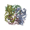
Mass: 663.425 Da / Num. of mol.: 4 / Source method: isolated from a natural source / Formula: C21H27N7O14P2 / Comment: NAD*YM
#3: Chemical
ChemComp-GOL / GLYCEROL / GLYCERIN / PROPANE-1,2,3-TRIOL / Glycerol


Mass: 92.094 Da / Num. of mol.: 12 / Source method: obtained synthetically / Formula: C3H8O3
#4: Water ChemComp-HOH / water / Water


Mass: 18.015 Da / Num. of mol.: 325 / Source method: isolated from a natural source / Formula: H2O

-
Experimental details

-
Experiment

ExperimentMethod: X-RAY DIFFRACTION / Number of used crystals: 1

-
Sample preparation

CrystalDensity Matthews: 2.43 Å3/Da / Density % sol: 49.3 % / Description: Plate or cube-shaped crystals
Crystal growTemperature: 293 K / Method: vapor diffusion, sitting drop / pH: 5.5 / Details: 0.1 M sodium citrate pH 5.5, 20% PEG 3000 / PH range: 5.0 - 6.0

-
Data collection

DiffractionMean temperature: 100 K
Diffraction sourceSource: SYNCHROTRON / Site: ALBA / Beamline: XALOC / Wavelength: 0.9795 Å
DetectorType: DECTRIS PILATUS3 6M / Detector: PIXEL / Date: May 5, 2015
RadiationMonochromator: Channel-cut Si(111) + KB focusing mirrors / Protocol: SINGLE WAVELENGTH / Monochromatic (M) / Laue (L): M / Scattering type: x-ray
Radiation wavelengthWavelength: 0.9795 Å / Relative weight: 1
ReflectionResolution: 2.1906→47.714 Å / Num. obs: 77410 / % possible obs: 99 % / Redundancy: 6.7 % / Biso Wilson estimate: 37.96 Å2 / CC1/2: 0.997 / Rmerge(I) obs: 0.1157 / Net I/σ(I): 9.46
Reflection shellResolution: 2.191→2.269 Å / Redundancy: 6.4 % / Rmerge(I) obs: 0.8224 / Mean I/σ(I) obs: 2.01 / % possible all: 90

-
Processing

Software
NameVersionClassification
XDSMarch 1, 2015data reduction
Aimless0.5.26data scaling
PHENIX1.9_1692refinement
PHASER2.6.1phasing
Coot0.8.2model building
RefinementMethod to determine structure: SAD / Resolution: 2.1906→47.714 Å / SU ML: 0.28 / Cross valid method: FREE R-VALUE / Phase error: 28.09
RfactorNum. reflection% reflectionSelection details
Rfree0.2266 3864 5 %Random selection
Rwork0.1773 ---
obs0.1797 77208 98.45 %-
Solvent computationShrinkage radii: 0.9 Å / VDW probe radii: 1.11 Å
Displacement parametersBiso mean: 37.96 Å2
Refinement stepCycle: LAST / Resolution: 2.1906→47.714 Å
ProteinNucleic acidLigandSolventTotal
Num. atoms10290 0 248 325 10863
Refine LS restraints
Refine-IDTypeDev idealNumber
X-RAY DIFFRACTIONf_bond_d0.00810798
X-RAY DIFFRACTIONf_angle_d1.09714679
X-RAY DIFFRACTIONf_dihedral_angle_d13.1443935
X-RAY DIFFRACTIONf_chiral_restr0.0441698
X-RAY DIFFRACTIONf_plane_restr0.0051877
LS refinement shell
Resolution (Å)Rfactor RfreeNum. reflection RfreeRfactor RworkNum. reflection RworkRefine-ID% reflection obs (%)
2.1906-2.21730.389940.35021782X-RAY DIFFRACTION68
2.2173-2.24540.33081420.27072688X-RAY DIFFRACTION99
2.2454-2.27490.27531360.26152584X-RAY DIFFRACTION99
2.2749-2.30610.29291400.25322664X-RAY DIFFRACTION100
2.3061-2.3390.29581390.25582628X-RAY DIFFRACTION100
2.339-2.37390.30961400.23792655X-RAY DIFFRACTION100
2.3739-2.4110.26911370.23932602X-RAY DIFFRACTION100
2.411-2.45060.30611410.22652682X-RAY DIFFRACTION100
2.4506-2.49280.28651390.23282639X-RAY DIFFRACTION100
2.4928-2.53810.30991400.22292645X-RAY DIFFRACTION100
2.5381-2.5870.29421390.22612644X-RAY DIFFRACTION100
2.587-2.63980.29561380.21542646X-RAY DIFFRACTION100
2.6398-2.69710.26271400.21642642X-RAY DIFFRACTION100
2.6971-2.75990.31321410.19952669X-RAY DIFFRACTION100
2.7599-2.82890.24791380.20232637X-RAY DIFFRACTION100
2.8289-2.90540.25661390.19942633X-RAY DIFFRACTION100
2.9054-2.99080.27811400.19972658X-RAY DIFFRACTION100
2.9908-3.08740.25271410.2042666X-RAY DIFFRACTION100
3.0874-3.19770.25121390.19232636X-RAY DIFFRACTION100
3.1977-3.32570.21331390.17972637X-RAY DIFFRACTION100
3.3257-3.4770.23591410.17242671X-RAY DIFFRACTION100
3.477-3.66030.21991400.17142657X-RAY DIFFRACTION100
3.6603-3.88950.18681400.15472656X-RAY DIFFRACTION100
3.8895-4.18960.18821410.14242685X-RAY DIFFRACTION100
4.1896-4.61090.16281390.12472633X-RAY DIFFRACTION100
4.6109-5.27740.16891410.13342683X-RAY DIFFRACTION100
5.2774-6.64610.20821410.15352670X-RAY DIFFRACTION100
6.6461-47.7140.17251390.14452652X-RAY DIFFRACTION97
Refinement TLS params.

Method: refined / Refine-ID: X-RAY DIFFRACTION

IDL112)L122)L132)L222)L232)L332)S11 (Å °)S12 (Å °)S13 (Å °)S21 (Å °)S22 (Å °)S23 (Å °)S31 (Å °)S32 (Å °)S33 (Å °)T112)T122)T132)T222)T232)T332)Origin x (Å)Origin y (Å)Origin z (Å)
11.33340.226-0.15852.65950.87661.3415-0.01790.06220.0139-0.36090.1265-0.2688-0.24980.2575-0.11180.4564-0.06060.08630.3702-0.0950.360719.5707-19.68344.6034
21.3093-0.3402-0.67762.2354-0.38631.5056-0.07910.0257-0.0312-0.18380.1847-0.22580.11490.1565-0.08740.3816-0.03060.05030.3392-0.11350.325715.9067-40.84265.7708
30.6443-0.22-0.12961.1224-0.21361.14210.02330.1169-0.0929-0.65070.04430.12080.2613-0.0141-0.050.3979-0.0275-0.00720.2632-0.00310.244-2.0949-37.076311.9499
41.1458-0.3455-0.52291.68330.37211.8765-0.0946-0.1746-0.17960.33350.1451-0.09450.32150.2568-0.03470.37840.0726-0.04420.292-0.01320.26569.3262-41.577823.5594
50.68850.2753-0.17482.0703-0.47061.8694-0.06390.42280.0475-0.64850.0230.3029-0.275-0.21420.03440.6781-0.0125-0.21560.53640.05470.5347-21.4421-15.9951-6.4148
60.5185-0.2086-0.33521.61340.14310.87550.0340.13920.0731-0.56930.06470.5064-0.2408-0.2125-0.08720.54490.0174-0.08560.33370.0650.4569-15.48592.841611.0309
71.26070.1002-0.52091.3295-1.29371.4533-0.07920.0460.2997-0.0252-0.01990.5849-0.0071-0.49190.1010.3018-0.020.02140.4512-0.0270.6372-32.2232-17.372233.8168
80.6989-0.5790.37691.33780.79921.83110.09-0.26260.04270.4877-0.21910.24350.3064-0.42370.12410.4483-0.09880.15740.49530.0360.5424-30.4989-34.246742.6315
90.58660.6348-0.03050.91410.1940.3206-0.05530.03010.06350.16370.11610.56760.2696-0.075-0.04320.4172-0.03250.05150.3270.05050.4526-12.7374-38.202429.6442
101.18180.1729-0.52730.9171-0.53141.41230.03240.1009-0.0033-0.1521-0.01040.28420.1952-0.2792-0.0070.3072-0.0932-0.02960.32230.00010.4501-20.4188-40.869219.1759
111.1382-0.38040.14441.96680.98962.4855-0.1351-0.13120.03060.46890.1352-0.18070.12070.289-0.02160.36410.0495-0.03980.3691-0.02790.249210.8975-20.014543.8678
121.1622-0.5696-0.29872.5789-0.59591.934-0.0516-0.44990.1150.66180.1823-0.1529-0.1450.1178-0.1220.44430.0472-0.05570.4138-0.07680.28165.3643-3.591951.312
130.8996-0.3405-0.05191.28470.38451.45990.0961-0.0890.11250.32910.05230.1417-0.25560.0004-0.1690.33940.00740.02750.26760.00190.263-4.07971.18430.1263
141.3665-0.3736-0.06241.9373-0.02761.4918-0.0172-0.01170.2111-0.36140.1296-0.3017-0.26220.2285-0.12350.3597-0.08590.05930.3049-0.05470.30649.32173.792625.6923
Refinement TLS group
IDRefine-IDRefine TLS-IDSelection details
1X-RAY DIFFRACTION1chain 'A' and (resid 15 through 119 )
2X-RAY DIFFRACTION2chain 'A' and (resid 120 through 183 )
3X-RAY DIFFRACTION3chain 'A' and (resid 184 through 267 )
4X-RAY DIFFRACTION4chain 'A' and (resid 268 through 355 )
5X-RAY DIFFRACTION5chain 'B' and (resid 16 through 136 )
6X-RAY DIFFRACTION6chain 'B' and (resid 137 through 355 )
7X-RAY DIFFRACTION7chain 'C' and (resid 15 through 93 )
8X-RAY DIFFRACTION8chain 'C' and (resid 94 through 167 )
9X-RAY DIFFRACTION9chain 'C' and (resid 168 through 244 )
10X-RAY DIFFRACTION10chain 'C' and (resid 245 through 356 )
11X-RAY DIFFRACTION11chain 'D' and (resid 16 through 92 )
12X-RAY DIFFRACTION12chain 'D' and (resid 93 through 167 )
13X-RAY DIFFRACTION13chain 'D' and (resid 168 through 267 )
14X-RAY DIFFRACTION14chain 'D' and (resid 268 through 355 )

+
About Yorodumi

-
News

-
Feb 9, 2022. New format data for meta-information of EMDB entries

New format data for meta-information of EMDB entries

  • Version 3 of the EMDB header file is now the official format.
  • The previous official version 1.9 will be removed from the archive.

Related info.:EMDB header

External links:wwPDB to switch to version 3 of the EMDB data model

-
Aug 12, 2020. Covid-19 info

Covid-19 info

URL: https://pdbj.org/emnavi/covid19.php

New page: Covid-19 featured information page in EM Navigator.

Related info.:Covid-19 info / Mar 5, 2020. Novel coronavirus structure data

+
Mar 5, 2020. Novel coronavirus structure data

Novel coronavirus structure data

Related info.:Yorodumi Speices / Aug 12, 2020. Covid-19 info

External links:COVID-19 featured content - PDBj / Molecule of the Month (242):Coronavirus Proteases

+
Jan 31, 2019. EMDB accession codes are about to change! (news from PDBe EMDB page)

EMDB accession codes are about to change! (news from PDBe EMDB page)

  • The allocation of 4 digits for EMDB accession codes will soon come to an end. Whilst these codes will remain in use, new EMDB accession codes will include an additional digit and will expand incrementally as the available range of codes is exhausted. The current 4-digit format prefixed with “EMD-” (i.e. EMD-XXXX) will advance to a 5-digit format (i.e. EMD-XXXXX), and so on. It is currently estimated that the 4-digit codes will be depleted around Spring 2019, at which point the 5-digit format will come into force.
  • The EM Navigator/Yorodumi systems omit the EMD- prefix.

Related info.:Q: What is EMD? / ID/Accession-code notation in Yorodumi/EM Navigator

External links:EMDB Accession Codes are Changing Soon! / Contact to PDBj

+
Jul 12, 2017. Major update of PDB

Major update of PDB

  • wwPDB released updated PDB data conforming to the new PDBx/mmCIF dictionary.
  • This is a major update changing the version number from 4 to 5, and with Remediation, in which all the entries are updated.
  • In this update, many items about electron microscopy experimental information are reorganized (e.g. em_software).
  • Now, EM Navigator and Yorodumi are based on the updated data.

External links:wwPDB Remediation / Enriched Model Files Conforming to OneDep Data Standards Now Available in the PDB FTP Archive

-
Yorodumi

Thousand views of thousand structures

  • Yorodumi is a browser for structure data from EMDB, PDB, SASBDB, etc.
  • This page is also the successor to EM Navigator detail page, and also detail information page/front-end page for Omokage search.
  • The word "yorodu" (or yorozu) is an old Japanese word meaning "ten thousand". "mi" (miru) is to see.

Related info.:EMDB / PDB / SASBDB / Comparison of 3 databanks / Yorodumi Search / Aug 31, 2016. New EM Navigator & Yorodumi / Yorodumi Papers / Jmol/JSmol / Function and homology information / Changes in new EM Navigator and Yorodumi

Read more