[English] 日本語
Yorodumi
- PDB-5lbm: The asymmetric tetrameric structure of the formaldehyde sensing t... -

+
Open data


ID or keywords:

Loading...

-
Basic information

Entry
Database: PDB / ID: 5lbm
TitleThe asymmetric tetrameric structure of the formaldehyde sensing transcriptional repressor FrmR from Escherichia coli
ComponentsTranscriptional repressor FrmR
KeywordsTRANSCRIPTION / CsoR/RcnR Escherichia coli FrmR methylene bridge
Function / homology
Function and homology information


regulation of DNA-templated transcription / DNA binding / metal ion binding / cytoplasm
Similarity search - Function
Metal-sensitive repressor, helix protomer / Metal-sensitive transcriptional repressor / Metal-sensitive repressor, helix protomer superfamily / Metal-sensitive transcriptional repressor / Methane Monooxygenase Hydroxylase; Chain G, domain 1 / Up-down Bundle / Mainly Alpha
Similarity search - Domain/homology
FORMYL GROUP / Transcriptional repressor FrmR
Similarity search - Component
Biological speciesEscherichia coli O157:H7 (bacteria)
MethodX-RAY DIFFRACTION / SYNCHROTRON / MOLECULAR REPLACEMENT / Resolution: 2.7 Å
AuthorsBisson, C. / Baker, P.J. / Green, J. / Chivers, P.T.
Funding support United Kingdom, 2items
OrganizationGrant numberCountry
Biotechnology and Biological Sciences Research CouncilBB/L008114/1 United Kingdom
Biotechnology and Biological Sciences Research CouncilBB/J014443/1 United Kingdom
CitationJournal: Sci Rep / Year: 2016
Title: The mechanism of a formaldehyde-sensing transcriptional regulator.
Authors: Denby, K.J. / Iwig, J. / Bisson, C. / Westwood, J. / Rolfe, M.D. / Sedelnikova, S.E. / Higgins, K. / Maroney, M.J. / Baker, P.J. / Chivers, P.T. / Green, J.
History
DepositionJun 16, 2016Deposition site: PDBE / Processing site: PDBE
Revision 1.0Dec 21, 2016Provider: repository / Type: Initial release
Revision 1.1Aug 30, 2017Group: Author supporting evidence / Category: pdbx_audit_support / Item: _pdbx_audit_support.funding_organization

-
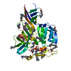
Structure visualization

Structure viewerMolecule:
MolmilJmol/JSmol

Downloads & links

-
Assembly

Deposited unit
A: Transcriptional repressor FrmR
B: Transcriptional repressor FrmR
C: Transcriptional repressor FrmR
D: Transcriptional repressor FrmR
hetero molecules


Theoretical massNumber of molelcules
Total (without water)41,7706
Polymers41,7104
Non-polymers602
Water0
1


  • Idetical with deposited unit
  • defined by software
TypeNameSymmetry operationNumber
identity operation1_555x,y,z1
Buried area10600 Å2
ΔGint-98 kcal/mol
Surface area17210 Å2
MethodPISA
Unit cell
Length a, b, c (Å)82.069, 82.069, 55.251
Angle α, β, γ (deg.)90.00, 90.00, 120.00
Int Tables number144
Space group name H-MP31

-
Components

#1: Protein
Transcriptional repressor FrmR


Mass: 10427.528 Da / Num. of mol.: 4
Source method: isolated from a genetically manipulated source
Source: (gene. exp.) Escherichia coli O157:H7 (bacteria) / Gene: frmR, Z0457, ECs0412 / Plasmid: pET22a / Production host: Escherichia coli (E. coli) / Strain (production host): BL21 / References: UniProt: Q8X5J3
#2: Chemical ChemComp-FOR / FORMYL GROUP / Aldehyde


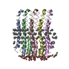
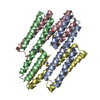
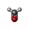
Mass: 30.026 Da / Num. of mol.: 2 / Source method: obtained synthetically / Formula: CH2O

-
Experimental details

-
Experiment

ExperimentMethod: X-RAY DIFFRACTION / Number of used crystals: 1

-
Sample preparation

CrystalDensity Matthews: 2.6 Å3/Da / Density % sol: 52.67 %
Crystal growTemperature: 290 K / Method: vapor diffusion, sitting drop / pH: 6.5
Details: 0.2 M MgCl2, 0.1 M Na cacodylate pH 6.5 and 31 % PEG 2000 Protein buffered in: 50mM Hepes pH 7.5 and 0.5 M NaCl

-
Data collection

DiffractionMean temperature: 100 K
Diffraction sourceSource: SYNCHROTRON / Site: Diamond / Beamline: I03 / Wavelength: 0.9763 Å
DetectorType: DECTRIS PILATUS 6M / Detector: PIXEL / Date: Feb 8, 2016
RadiationProtocol: SINGLE WAVELENGTH / Monochromatic (M) / Laue (L): M / Scattering type: x-ray
Radiation wavelengthWavelength: 0.9763 Å / Relative weight: 1
ReflectionResolution: 2.7→55.25 Å / Num. obs: 11450 / % possible obs: 100 % / Redundancy: 5.3 % / Rpim(I) all: 0.038 / Net I/σ(I): 12.2
Reflection shellResolution: 2.7→2.83 Å / Redundancy: 5.4 % / Rmerge(I) obs: 0.499 / Mean I/σ(I) obs: 2.2 / Rpim(I) all: 0.499 / % possible all: 100

-
Processing

Software
NameVersionClassification
REFMAC5.8.0151refinement
XDSdata reduction
xia2data reduction
Aimlessdata scaling
PHASERphasing
BUCCANEERphasing
RefinementMethod to determine structure: MOLECULAR REPLACEMENT / Resolution: 2.7→55.25 Å / Cor.coef. Fo:Fc: 0.957 / Cor.coef. Fo:Fc free: 0.94 / SU B: 14.808 / SU ML: 0.301 / Cross valid method: THROUGHOUT / ESU R Free: 0.368 / Details: HYDROGENS HAVE BEEN ADDED IN THE RIDING POSITIONS
RfactorNum. reflection% reflectionSelection details
Rfree0.26628 567 5 %RANDOM
Rwork0.20879 ---
obs0.2117 10865 99.99 %-
Solvent computationIon probe radii: 0.8 Å / Shrinkage radii: 0.8 Å / VDW probe radii: 1.2 Å
Displacement parametersBiso mean: 79.267 Å2
Baniso -1Baniso -2Baniso -3
1-0.39 Å20.19 Å20 Å2
2--0.39 Å20 Å2
3----1.26 Å2
Refinement stepCycle: 1 / Resolution: 2.7→55.25 Å
ProteinNucleic acidLigandSolventTotal
Num. atoms2744 0 2 0 2746
Refine LS restraints
Refine-IDTypeDev idealDev ideal targetNumber
X-RAY DIFFRACTIONr_bond_refined_d0.0120.0192766
X-RAY DIFFRACTIONr_bond_other_d0.0020.022748
X-RAY DIFFRACTIONr_angle_refined_deg1.5981.9793708
X-RAY DIFFRACTIONr_angle_other_deg0.9823.0016270
X-RAY DIFFRACTIONr_dihedral_angle_1_deg4.6885342
X-RAY DIFFRACTIONr_dihedral_angle_2_deg34.68622.778144
X-RAY DIFFRACTIONr_dihedral_angle_3_deg15.80415524
X-RAY DIFFRACTIONr_dihedral_angle_4_deg15.7641544
X-RAY DIFFRACTIONr_chiral_restr0.0850.2428
X-RAY DIFFRACTIONr_gen_planes_refined0.0050.023132
X-RAY DIFFRACTIONr_gen_planes_other0.0020.02632
X-RAY DIFFRACTIONr_nbd_refined
X-RAY DIFFRACTIONr_nbd_other
X-RAY DIFFRACTIONr_nbtor_refined
X-RAY DIFFRACTIONr_nbtor_other
X-RAY DIFFRACTIONr_xyhbond_nbd_refined
X-RAY DIFFRACTIONr_xyhbond_nbd_other
X-RAY DIFFRACTIONr_metal_ion_refined
X-RAY DIFFRACTIONr_metal_ion_other
X-RAY DIFFRACTIONr_symmetry_vdw_refined
X-RAY DIFFRACTIONr_symmetry_vdw_other
X-RAY DIFFRACTIONr_symmetry_hbond_refined
X-RAY DIFFRACTIONr_symmetry_hbond_other
X-RAY DIFFRACTIONr_symmetry_metal_ion_refined
X-RAY DIFFRACTIONr_symmetry_metal_ion_other
X-RAY DIFFRACTIONr_mcbond_it3.5287.6181382
X-RAY DIFFRACTIONr_mcbond_other3.5277.6181381
X-RAY DIFFRACTIONr_mcangle_it5.55411.411718
X-RAY DIFFRACTIONr_mcangle_other5.55311.411719
X-RAY DIFFRACTIONr_scbond_it3.7198.3091384
X-RAY DIFFRACTIONr_scbond_other3.7198.311384
X-RAY DIFFRACTIONr_scangle_it
X-RAY DIFFRACTIONr_scangle_other6.16712.2331990
X-RAY DIFFRACTIONr_long_range_B_refined8.49190.5563084
X-RAY DIFFRACTIONr_long_range_B_other8.4990.5693085
X-RAY DIFFRACTIONr_rigid_bond_restr
X-RAY DIFFRACTIONr_sphericity_free
X-RAY DIFFRACTIONr_sphericity_bonded
LS refinement shellResolution: 2.701→2.771 Å / Total num. of bins used: 20
RfactorNum. reflection% reflection
Rfree0.385 51 -
Rwork0.298 813 -
obs--100 %

+
About Yorodumi

-
News

-
Feb 9, 2022. New format data for meta-information of EMDB entries

New format data for meta-information of EMDB entries

  • Version 3 of the EMDB header file is now the official format.
  • The previous official version 1.9 will be removed from the archive.

Related info.:EMDB header

External links:wwPDB to switch to version 3 of the EMDB data model

-
Aug 12, 2020. Covid-19 info

Covid-19 info

URL: https://pdbj.org/emnavi/covid19.php

New page: Covid-19 featured information page in EM Navigator.

Related info.:Covid-19 info / Mar 5, 2020. Novel coronavirus structure data

+
Mar 5, 2020. Novel coronavirus structure data

Novel coronavirus structure data

Related info.:Yorodumi Speices / Aug 12, 2020. Covid-19 info

External links:COVID-19 featured content - PDBj / Molecule of the Month (242):Coronavirus Proteases

+
Jan 31, 2019. EMDB accession codes are about to change! (news from PDBe EMDB page)

EMDB accession codes are about to change! (news from PDBe EMDB page)

  • The allocation of 4 digits for EMDB accession codes will soon come to an end. Whilst these codes will remain in use, new EMDB accession codes will include an additional digit and will expand incrementally as the available range of codes is exhausted. The current 4-digit format prefixed with “EMD-” (i.e. EMD-XXXX) will advance to a 5-digit format (i.e. EMD-XXXXX), and so on. It is currently estimated that the 4-digit codes will be depleted around Spring 2019, at which point the 5-digit format will come into force.
  • The EM Navigator/Yorodumi systems omit the EMD- prefix.

Related info.:Q: What is EMD? / ID/Accession-code notation in Yorodumi/EM Navigator

External links:EMDB Accession Codes are Changing Soon! / Contact to PDBj

+
Jul 12, 2017. Major update of PDB

Major update of PDB

  • wwPDB released updated PDB data conforming to the new PDBx/mmCIF dictionary.
  • This is a major update changing the version number from 4 to 5, and with Remediation, in which all the entries are updated.
  • In this update, many items about electron microscopy experimental information are reorganized (e.g. em_software).
  • Now, EM Navigator and Yorodumi are based on the updated data.

External links:wwPDB Remediation / Enriched Model Files Conforming to OneDep Data Standards Now Available in the PDB FTP Archive

-
Yorodumi

Thousand views of thousand structures

  • Yorodumi is a browser for structure data from EMDB, PDB, SASBDB, etc.
  • This page is also the successor to EM Navigator detail page, and also detail information page/front-end page for Omokage search.
  • The word "yorodu" (or yorozu) is an old Japanese word meaning "ten thousand". "mi" (miru) is to see.

Related info.:EMDB / PDB / SASBDB / Comparison of 3 databanks / Yorodumi Search / Aug 31, 2016. New EM Navigator & Yorodumi / Yorodumi Papers / Jmol/JSmol / Function and homology information / Changes in new EM Navigator and Yorodumi

Read more