 Movie
Movie Controller


+ Open data

- Basic information
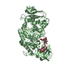
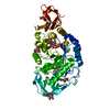
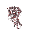
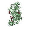
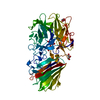
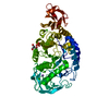
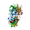
| Entry | Database: PDB / ID: 5lae | ||||||
|---|---|---|---|---|---|---|---|
| Title | Crystal structure of murine N1-acetylpolyamine oxidase | ||||||
 Components Components | Peroxisomal N(1)-acetyl-spermine/spermidine oxidase,Peroxisomal N(1)-acetyl-spermine/spermidine oxidase | ||||||
 Keywords Keywords | OXIDOREDUCTASE / flavin amine oxidase | ||||||
| Function / homology |  Function and homology information Function and homology informationN1-acetylpolyamine oxidase / PAOs oxidise polyamines to amines / positive regulation of spermidine biosynthetic process / Interconversion of polyamines / putrescine biosynthetic process / N(1)-acetylpolyamine oxidase activity / polyamine oxidase activity / putrescine catabolic process / polyamine catabolic process / spermine catabolic process ...N1-acetylpolyamine oxidase / PAOs oxidise polyamines to amines / positive regulation of spermidine biosynthetic process / Interconversion of polyamines / putrescine biosynthetic process / N(1)-acetylpolyamine oxidase activity / polyamine oxidase activity / putrescine catabolic process / polyamine catabolic process / spermine catabolic process / spermidine catabolic process / Peroxisomal protein import / peroxisomal matrix / cytoplasm Similarity search - Function | ||||||
| Biological species |  | ||||||
| Method |  X-RAY DIFFRACTION / SYNCHROTRON / MOLECULAR REPLACEMENT / Resolution: 1.85 Å X-RAY DIFFRACTION / SYNCHROTRON / MOLECULAR REPLACEMENT / Resolution: 1.85 Å | ||||||
 Authors Authors | Sjogren, T. / Aagaard, A. / Snijder, A. / Barlind, L. | ||||||
 Citation Citation | Journal: Biochemistry / Year: 2017 Title: The Structure of Murine N(1)-Acetylspermine Oxidase Reveals Molecular Details of Vertebrate Polyamine Catabolism. Authors: Sjogren, T. / Wassvik, C.M. / Snijder, A. / Aagaard, A. / Kumanomidou, T. / Barlind, L. / Kaminski, T.P. / Kashima, A. / Yokota, T. / Fjellstrom, O. | ||||||
| History |
|
- Structure visualization
| Structure viewer | Molecule: MolmilJmol/JSmol |
|---|
- Downloads & links
-Download
| PDBx/mmCIF format | 5lae.cif.gz | 113.3 KB | Display | PDBx/mmCIF format |
|---|---|---|---|---|
| PDB format | pdb5lae.ent.gz | 83.9 KB | Display | PDB format |
| PDBx/mmJSON format | 5lae.json.gz | Tree view | PDBx/mmJSON format | |
| Others |  Other downloads Other downloads |
-Validation report
| Arichive directory | https://data.pdbj.org/pub/pdb/validation_reports/la/5laeftp://data.pdbj.org/pub/pdb/validation_reports/la/5lae | HTTPS FTP |
|---|
-Related structure data
| Related structure data | 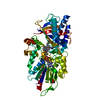 5lfoC 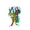 5lgbC 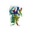 5mbxC 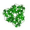 1bq5S S: Starting model for refinement C: citing same article ( |
|---|---|
| Similar structure data |


-Links
 PDBj
PDBj

- Assembly
| Deposited unit | 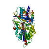
| ||||||||
|---|---|---|---|---|---|---|---|---|---|
| 1 |
| ||||||||
| Unit cell |
|
-Components
| #1: Protein | Mass: 54634.645 Da / Num. of mol.: 1 Source method: isolated from a genetically manipulated source Details: The sequence includes 3 aa N-terminal deletion and a loop deletion where 6 residues (451-457) are replaced with a single glycine residue. Source: (gene. exp.)  | ||
|---|---|---|---|
| #2: Chemical | ChemComp-FAD /   Mass: 785.550 Da / Num. of mol.: 1 / Source method: obtained synthetically / Formula: C27H33N9O15P2 / Comment: FAD*YM Mass: 785.550 Da / Num. of mol.: 1 / Source method: obtained synthetically / Formula: C27H33N9O15P2 / Comment: FAD*YM | ||
| #3: Chemical |   Mass: 92.094 Da / Num. of mol.: 2 / Source method: obtained synthetically / Formula: C3H8O3 Mass: 92.094 Da / Num. of mol.: 2 / Source method: obtained synthetically / Formula: C3H8O3#4: Water | ChemComp-HOH / |  Mass: 18.015 Da / Num. of mol.: 287 / Source method: isolated from a natural source / Formula: H2O Mass: 18.015 Da / Num. of mol.: 287 / Source method: isolated from a natural source / Formula: H2O |
-Experimental details
-Experiment
| Experiment | Method: X-RAY DIFFRACTION / Number of used crystals: 1 |
|---|
- Sample preparation
| Crystal | Density Matthews: 2.17 Å3/Da / Density % sol: 43.2 % |
|---|---|
| Crystal grow | Temperature: 293 K / Method: vapor diffusion, hanging drop / pH: 8 Details: Protein at 23mg/ml in 20 mM HEPES, pH 7.0, 100 mM NaCl, 5% (v/v) glycerol and 2.5 mM TCEP was equilibrated against 2.2 M (NH4)2SO4, 0.2 M NaSCN, 0.1 M Tris pH 8 |
-Data collection
| Diffraction | Mean temperature: 100 K |
|---|---|
| Diffraction source | Source: SYNCHROTRON / Site: ESRF  / Beamline: ID23-1 / Wavelength: 0.972 Å / Beamline: ID23-1 / Wavelength: 0.972 Å |
| Detector | Type: DECTRIS PILATUS 6M / Detector: PIXEL / Date: Jan 26, 2015 |
| Radiation | Monochromator: Single crystal / Protocol: SINGLE WAVELENGTH / Monochromatic (M) / Laue (L): M / Scattering type: x-ray |
| Radiation wavelength | Wavelength: 0.972 Å / Relative weight: 1 |
| Reflection | Resolution: 1.85→49 Å / Num. obs: 40245 / % possible obs: 99.2 % / Redundancy: 9.8 % / Biso Wilson estimate: 26.96 Å2 / CC1/2: 0.999 / Rmerge(I) obs: 0.109 / Net I/σ(I): 14.9 |
| Reflection shell | Resolution: 1.85→1.89 Å / Redundancy: 6.5 % / Rmerge(I) obs: 0.87 / Mean I/σ(I) obs: 1.9 / % possible all: 95 |
- Processing
| Software |
| ||||||||||||||||||||||||||||||||||||||||||||||||||||||||||||||||||||||||||||||||||||||||||||||||||||||||||||||||||
|---|---|---|---|---|---|---|---|---|---|---|---|---|---|---|---|---|---|---|---|---|---|---|---|---|---|---|---|---|---|---|---|---|---|---|---|---|---|---|---|---|---|---|---|---|---|---|---|---|---|---|---|---|---|---|---|---|---|---|---|---|---|---|---|---|---|---|---|---|---|---|---|---|---|---|---|---|---|---|---|---|---|---|---|---|---|---|---|---|---|---|---|---|---|---|---|---|---|---|---|---|---|---|---|---|---|---|---|---|---|---|---|---|---|---|---|
| Refinement | Method to determine structure: MOLECULAR REPLACEMENT Starting model: 1BQ5 Resolution: 1.85→49 Å / Cor.coef. Fo:Fc: 0.954 / Cor.coef. Fo:Fc free: 0.947 / SU R Cruickshank DPI: 0.131 / Cross valid method: THROUGHOUT / σ(F): 0 / SU R Blow DPI: 0.134 / SU Rfree Blow DPI: 0.121 / SU Rfree Cruickshank DPI: 0.121
| ||||||||||||||||||||||||||||||||||||||||||||||||||||||||||||||||||||||||||||||||||||||||||||||||||||||||||||||||||
| Displacement parameters | Biso mean: 31.58 Å2
| ||||||||||||||||||||||||||||||||||||||||||||||||||||||||||||||||||||||||||||||||||||||||||||||||||||||||||||||||||
| Refine analyze | Luzzati coordinate error obs: 0.21 Å | ||||||||||||||||||||||||||||||||||||||||||||||||||||||||||||||||||||||||||||||||||||||||||||||||||||||||||||||||||
| Refinement step | Cycle: LAST / Resolution: 1.85→49 Å
| ||||||||||||||||||||||||||||||||||||||||||||||||||||||||||||||||||||||||||||||||||||||||||||||||||||||||||||||||||
| Refine LS restraints |
| ||||||||||||||||||||||||||||||||||||||||||||||||||||||||||||||||||||||||||||||||||||||||||||||||||||||||||||||||||
| LS refinement shell | Resolution: 1.85→1.9 Å / Rfactor Rfree error: 0 / Total num. of bins used: 20
|
