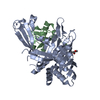
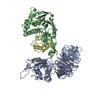
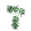
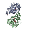
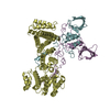
STRUCTURAL PROTEIN / DUB-Activator complex USP1 paralog
Function / homology
Function and homology information
regulation of protein monoubiquitination / Signaling by cytosolic PDGFRA and PDGFRB fusion proteins / deubiquitinase activator activity / hypothalamus gonadotrophin-releasing hormone neuron development / female meiosis I / skeletal system morphogenesis / positive regulation of protein monoubiquitination / fat pad development / mitochondrion transport along microtubule / skin development ...regulation of protein monoubiquitination / Signaling by cytosolic PDGFRA and PDGFRB fusion proteins / deubiquitinase activator activity / hypothalamus gonadotrophin-releasing hormone neuron development / female meiosis I / skeletal system morphogenesis / positive regulation of protein monoubiquitination / fat pad development / mitochondrion transport along microtubule / skin development / seminiferous tubule development / female gonad development / male meiosis I / homeostasis of number of cells / embryonic organ development / single fertilization / positive regulation of intrinsic apoptotic signaling pathway by p53 class mediator / protein deubiquitination / positive regulation of double-strand break repair via homologous recombination / energy homeostasis / neuron projection morphogenesis / regulation of proteasomal protein catabolic process / Maturation of protein E / Maturation of protein E / ER Quality Control Compartment (ERQC) / Myoclonic epilepsy of Lafora / FLT3 signaling by CBL mutants / IRAK2 mediated activation of TAK1 complex / Alpha-protein kinase 1 signaling pathway / Glycogen synthesis / IRAK1 recruits IKK complex / IRAK1 recruits IKK complex upon TLR7/8 or 9 stimulation / Prevention of phagosomal-lysosomal fusion / Endosomal Sorting Complex Required For Transport (ESCRT) / Membrane binding and targetting of GAG proteins / Regulation of TBK1, IKKε (IKBKE)-mediated activation of IRF3, IRF7 / Negative regulation of FLT3 / PTK6 Regulates RTKs and Their Effectors AKT1 and DOK1 / Regulation of TBK1, IKKε-mediated activation of IRF3, IRF7 upon TLR3 ligation / IRAK2 mediated activation of TAK1 complex upon TLR7/8 or 9 stimulation / Constitutive Signaling by NOTCH1 HD Domain Mutants / NOTCH2 Activation and Transmission of Signal to the Nucleus / TICAM1,TRAF6-dependent induction of TAK1 complex / TICAM1-dependent activation of IRF3/IRF7 / APC/C:Cdc20 mediated degradation of Cyclin B / regulation of neuron apoptotic process / Downregulation of ERBB4 signaling / APC-Cdc20 mediated degradation of Nek2A / Regulation of FZD by ubiquitination / p75NTR recruits signalling complexes / InlA-mediated entry of Listeria monocytogenes into host cells / TRAF6 mediated IRF7 activation in TLR7/8 or 9 signaling / positive regulation of epithelial cell proliferation / NF-kB is activated and signals survival / TRAF6-mediated induction of TAK1 complex within TLR4 complex / Regulation of pyruvate metabolism / Pexophagy / Downregulation of ERBB2:ERBB3 signaling / Regulation of innate immune responses to cytosolic DNA / NRIF signals cell death from the nucleus / Regulation of PTEN localization / positive regulation of protein ubiquitination / VLDLR internalisation and degradation / Activated NOTCH1 Transmits Signal to the Nucleus / Synthesis of active ubiquitin: roles of E1 and E2 enzymes / Translesion synthesis by REV1 / TICAM1, RIP1-mediated IKK complex recruitment / Regulation of BACH1 activity / Translesion synthesis by POLK / JNK (c-Jun kinases) phosphorylation and activation mediated by activated human TAK1 / InlB-mediated entry of Listeria monocytogenes into host cell / MAP3K8 (TPL2)-dependent MAPK1/3 activation / Activation of IRF3, IRF7 mediated by TBK1, IKKε (IKBKE) / Downregulation of TGF-beta receptor signaling / Translesion synthesis by POLI / Josephin domain DUBs / Gap-filling DNA repair synthesis and ligation in GG-NER / IKK complex recruitment mediated by RIP1 / positive regulation of receptor signaling pathway via JAK-STAT / PINK1-PRKN Mediated Mitophagy / regulation of mitochondrial membrane potential / TGF-beta receptor signaling in EMT (epithelial to mesenchymal transition) / TNFR1-induced NF-kappa-B signaling pathway / Regulation of activated PAK-2p34 by proteasome mediated degradation / TCF dependent signaling in response to WNT / Regulation of NF-kappa B signaling / activated TAK1 mediates p38 MAPK activation / ubiquitin binding / Autodegradation of Cdh1 by Cdh1:APC/C / APC/C:Cdc20 mediated degradation of Securin / NOTCH3 Activation and Transmission of Signal to the Nucleus / Regulation of signaling by CBL / Negative regulators of DDX58/IFIH1 signaling / N-glycan trimming in the ER and Calnexin/Calreticulin cycle / Asymmetric localization of PCP proteins / Fanconi Anemia Pathway / Negative regulation of FGFR3 signaling / Ubiquitin-dependent degradation of Cyclin D / Peroxisomal protein import / Deactivation of the beta-catenin transactivating complex Similarity search - Function
Mass: 65194.832 Da / Num. of mol.: 1 Source method: isolated from a genetically manipulated source Source: (gene. exp.) Homo sapiens (human) / Gene: WDR48, KIAA1449, UAF1 / Production host: Spodoptera frugiperda (fall armyworm) / References: UniProt: Q8TAF3
#3: Protein
Polyubiquitin-B
Mass: 8519.778 Da / Num. of mol.: 1 Source method: isolated from a genetically manipulated source Details: Recombinant expression followed by chemical synthesis Source: (gene. exp.) Homo sapiens (human) / Gene: UBB / Production host: Escherichia coli (E. coli) / References: UniProt: P0CG47
Mass: 57.094 Da / Num. of mol.: 1 / Source method: obtained synthetically / Formula: C3H7N Details: Recombinant expression followed by chemical synthesis Source: (gene. exp.) Homo sapiens (human) / Production host: Escherichia coli (E. coli)
 Movie
Movie Controller


 Open data
 Basic information
 Components
 Keywords
 Function and homology information
 Homo sapiens (human)
 X-RAY DIFFRACTION /
  Authors
 Netherlands, 3items
  Citation
 Structure visualization
 Downloads & links
 Other downloads
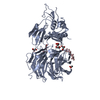
 PDBj
PDBj


 Assembly

 Spodoptera frugiperda (fall armyworm) / References: UniProt: O75317, ubiquitinyl hydrolase 1


 Mass: 65.409 Da / Num. of mol.: 1 / Source method: obtained synthetically / Formula: Zn
 Mass: 92.094 Da / Num. of mol.: 1 / Source method: obtained synthetically / Formula: C3H8O3
 Mass: 57.094 Da / Num. of mol.: 1 / Source method: obtained synthetically / Formula: C3H7N
 Sample preparation
 / Beamline: X06DA / Wavelength: 1 Å
 Processing